

Digital Twins for Personalized Cardiovascular Health
twinHeart builds digital twins of the human thorax to understand where heart vibration signals come from and what they can reveal. Starting from 4D CT or MR images, we reconstruct the heart, lungs, ribcage, muscles, and fat, track the motion of the heart wall through the cardiac cycle, and use finite element modeling to simulate how that motion travels to the chest surface, producing a simulated seismocardiogram (SCG). These models let us test how SCG signals change with tissue properties and myocardial motion, which is difficult to vary in people.
We also estimate myocardial strain, an early marker of heart dysfunction, from 4D CT and MR images using optical flow, deep learning, and finite element modeling. Our publications include a practical guide to finite element modeling of SCG and a review of deep learning for cardiac wall motion analysis.
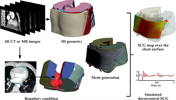
Develop digital twins of the human thorax to facilitate the design, testing, and validation of innovative diagnostic and therapeutic methods for a wide range of cardiovascular conditions

Principal Investigator

In this podcast episode, we break down the goals and methods of this project in a lay language. Alternatively, you can read our review article below.
Read Article

We design intelligent medical decision-making solutions to improve health equity.
111 Research Dr., Rm. C346, Bethlehem, PA 18015
+1 (610) 758-4096
taebilab@gmail.com
© TaebiLab. All Rights Reserved.
Adapted from a design by HTML Codex