
Taebi Lab Logo
Lehigh University Logo
-
Publication Stats

* Current and former lab members are underlined.
Publication Thumbnail

Monfared, M., Kakavand, B., Taebi, A. (2026).
Deep Learning for Cardiac Wall Motion Analysis: A Review of Methods, Challenges, and Clinical Applications.
Annals of Biomedical Engineering.
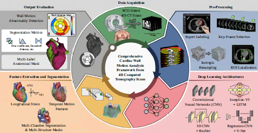
Abstract: Abnormalities in cardiac wall motion are strong predictors of cardiovascular risk, making their accurate detection essential for early diagnosis and effective clinical management. Traditional imaging modalities such as echocardiography, magnetic resonance imaging (MRI), and computed tomography (CT) provide valuable insights but face limitations related to accessibility, cost, and the complexity of spatiotemporal analysis. Recent advances in machine learning (ML), particularly deep learning (DL), have enabled automated extraction of spatial and temporal features from medical imaging. They improved accuracy in segmentation, motion estimation, and detection of regional wall motion abnormalities. This paper reviews state-of-the-art methods for predicting cardiac wall motion, with emphasis on DL applications across echocardiography, 4D CT, and cine MRI datasets. Representative studies demonstrate the potential of convolutional neural networks, recurrent neural networks, and transformers to achieve performance comparable to expert clinicians, while also highlighting challenges such as data scarcity, model interpretability, and limited external validation. Addressing these issues will be critical for translating ML-based approaches into routine practice, and integration of advanced imaging with robust ML frameworks helps in developing a reliable cardiac wall motion simulators for personalized treatment planning and improved cardiovascular care.
Publication Thumbnail

Roncali, E., Taebi, A. (2026).
Computational Fluid Dynamics Simulations to Inform Cancer Therapeutics.
Annual Review of Biomedical Engineering, 28: 515-38.
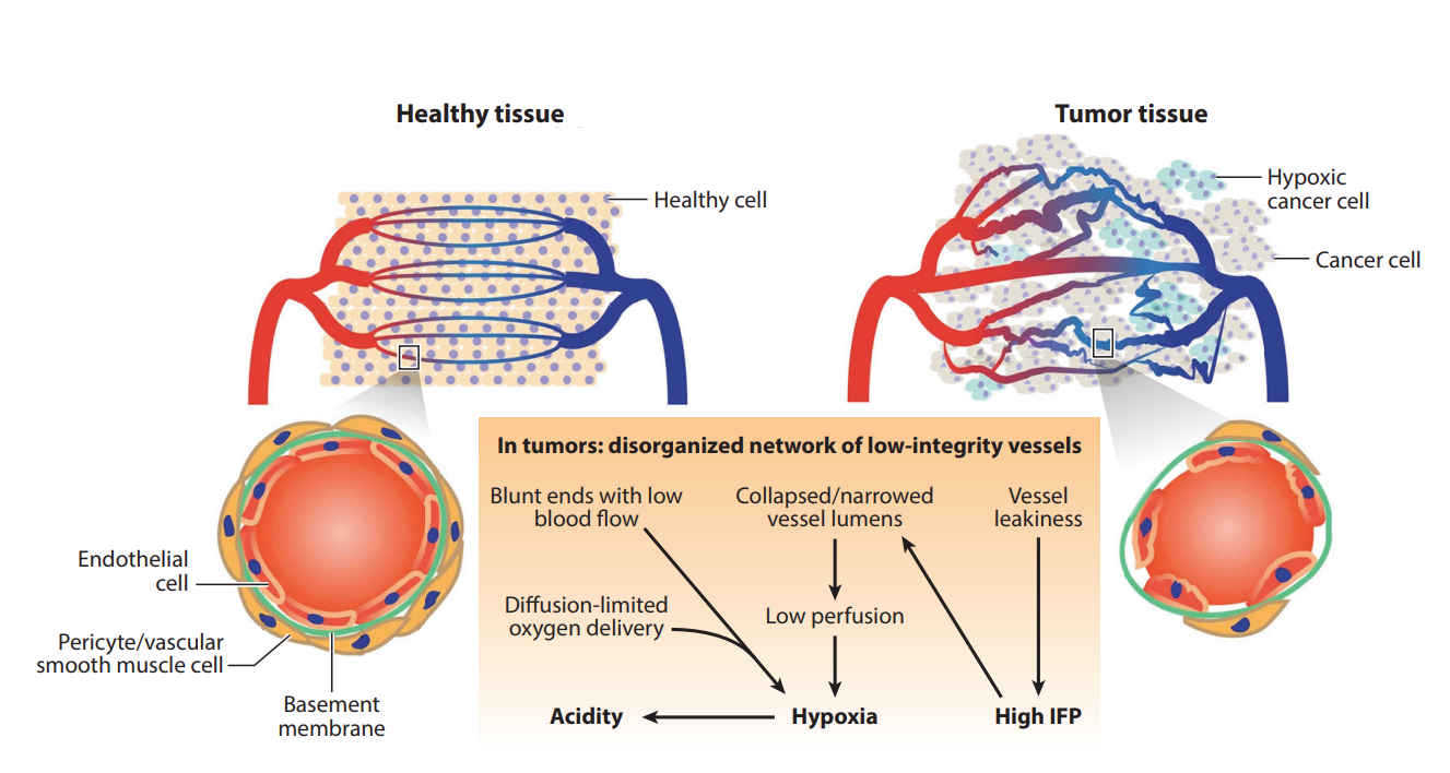
Abstract: Cancer therapies such as chemotherapy, radiopharmaceutical therapy, and transarterial embolization rely on effective drug or radiation delivery through the bloodstream. Understanding how various drugs and particles, which form and size span multiple scales, are transported through blood and tissue is essential for optimizing treatment. Computational fluid dynamics (CFD) is a powerful tool to simulate blood flow and drug transport, solving flow governing equations under biologically realistic conditions. This review explores CFD applications in cancer therapy, focusing on transarterial embolization, tumor perfusion, and organ-on-a-chip systems. In radioembolization, CFD can predict microsphere transport and dose distribution to spare vital functions. Tumor perfusion modeling and organ-on-a-chip systems benefit from CFD by replicating vascular dynamics and drug dispersion. Despite its versatility and established mechanical principles, CFD faces challenges, including the need for patient-specific data, computational demands, and multiscale modeling. This review highlights opportunities for integrating CFD with imaging modalities and artificial intelligence tools to overcome these barriers and advance personalized cancer treatment.
Publication Thumbnail

Rahman, M.M., Taebi, A. (2026).
Heart Rate Monitoring from Smartphone Neck Videos Using Remote Photoplethysmography.
ASME Journal of Engineering and Science in Medical Diagnostics and Therapy, 9(2): 021009.
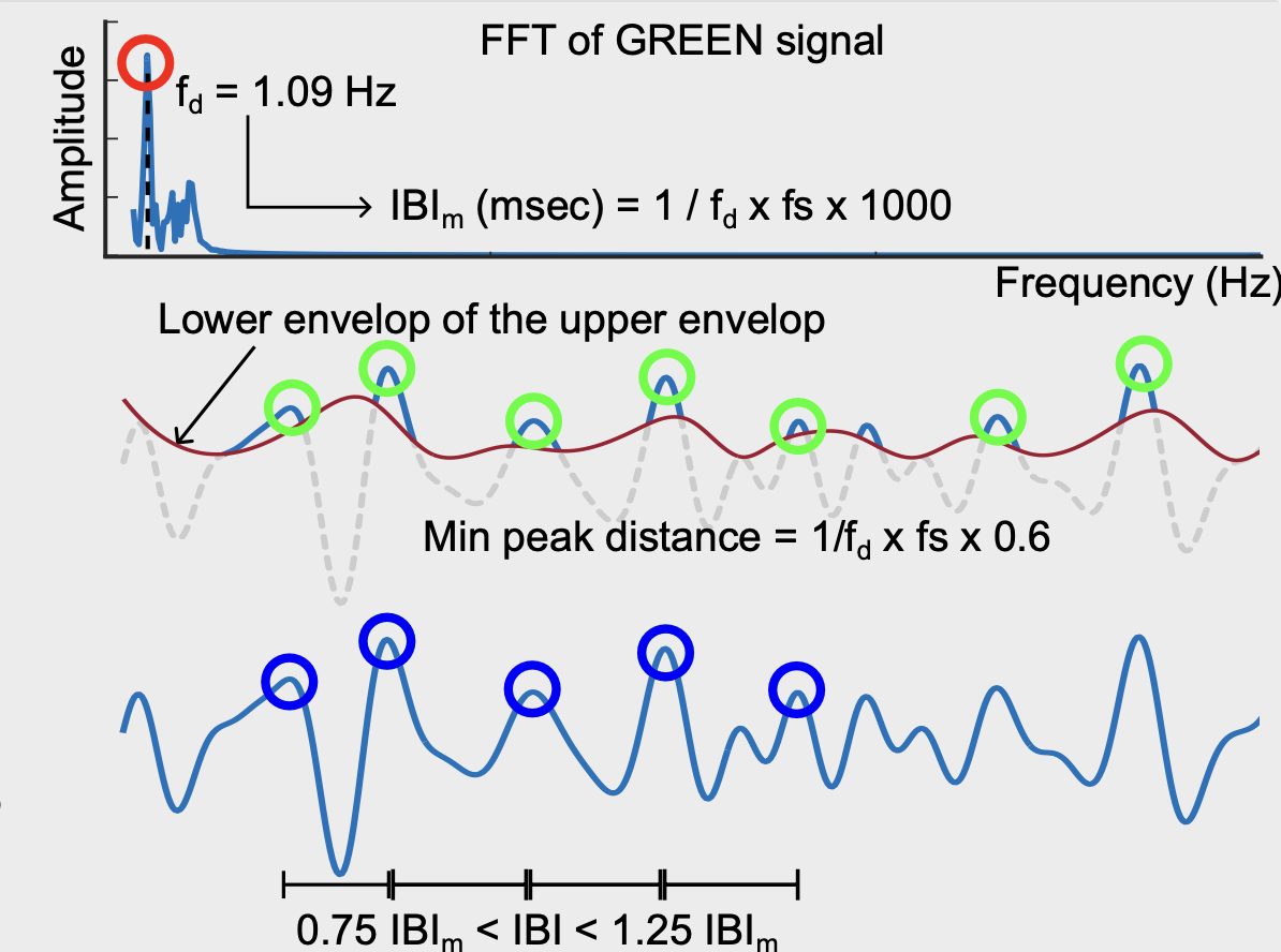
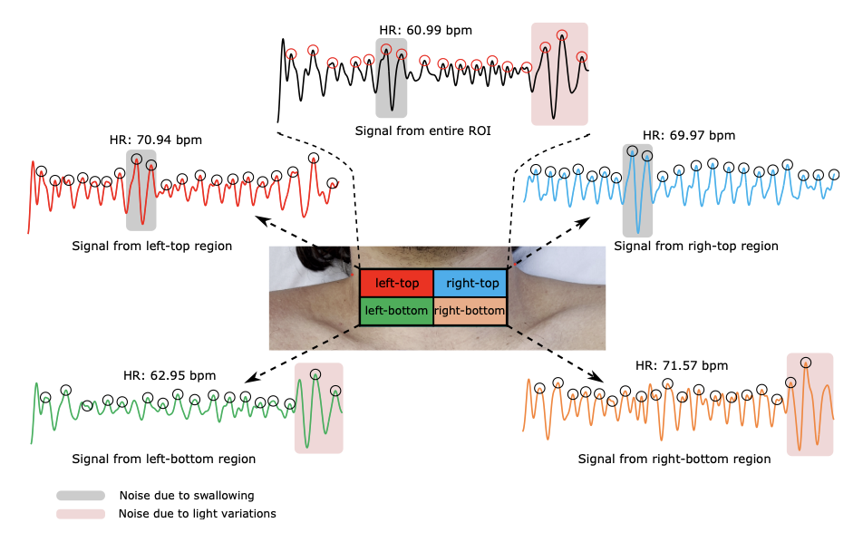
Abstract: Remote photoplethysmography (rPPG) enables contactless estimation of physiological signals from skin videos, offering a promising solution for unobtrusive cardiovascular monitoring. While most rPPG studies have focused on facial regions, privacy concerns limit their broader applicability. In this study, we explore the neck area as an alternative region of interest (ROI) for rPPG-based heart rate (HR) estimation, leveraging its proximity to major blood vessels. Video recordings of the neck of 15 adult subjects were recorded and processed using six rPPG methods: GREEN, CHROM, POS, OMIT, ICA, and LGI. HR estimates derived from each method were compared against those calculated from a gold-standard fingertip PPG using Bland–Altman and correlation analysis. Among all methods, GREEN method demonstrated the best agreement, with a bias of −0.82 bpm and limits of agreement (LoA) of −8.86–7.22 bpm, followed closely by POS (bias = −0.10 bpm; LoA = −8.50–8.30 bpm). Both methods also exhibited strong linear correlation with reference HR (r = 0.95), indicating excellent consistency. Spatial analysis of the neck region identified localized artifacts due to swallowing, light illumination, and vascular asymmetry as potential sources of signal degradation in certain cases. These findings demonstrate the viability of neck-based rPPG for HR monitoring while highlighting the importance of method selection and region-specific optimization. The study provides valuable insights for developing robust, privacy-conscious rPPG systems in clinical and remote healthcare applications.
Publication Thumbnail

Monfared, M., Kakavand, B., Gamage, P.T., Taebi, A. (2025).
Digital twin-based investigation of seismocardiogram sensitivity to tissue mechanics and myocardial motion.
ASME Journal of Biomechanical Engineering, 147(12): 121007.​

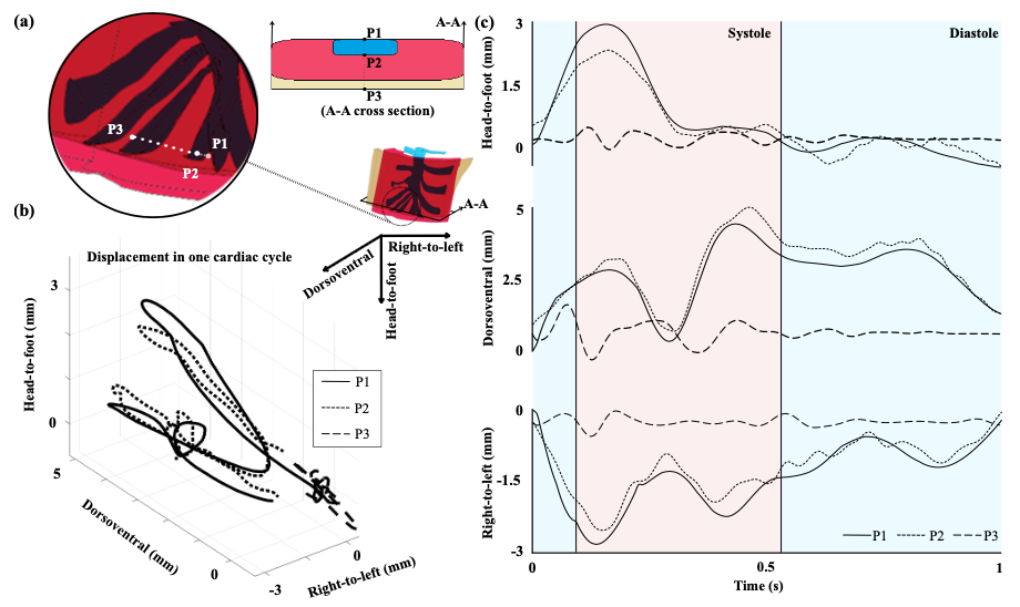
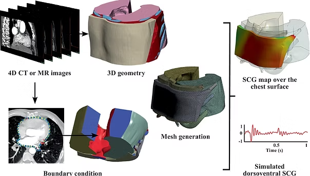
Abstract: Cardiovascular diseases remain the leading cause of mortality worldwide, underscoring the need for improved diagnostic tools. Seismocardiography (SCG), a noninvasive technique that records chest surface vibrations generated by cardiac activity, holds promise for such applications. However, the mechanistic origins of SCG waveforms, particularly under varying physiological conditions, remain insufficiently understood. This study presents a finite element modeling approach to simulate SCG signals by tracking the propagation of cardiac wall motion to the chest surface. The computational model, constructed from 4D computed tomography (CT) scans of healthy adult subjects, incorporates the lungs, ribcage, muscles, and adipose tissue. Cardiac displacement boundary conditions were extracted using the Lucas-Kanade algorithm, and elastic properties were assigned to different tissues. The simulated SCG signals in the dorsoventral direction were compared to realistic SCG recordings, showing consistency in waveform morphology. Key cardiac events, such as mitral valve closure, aortic valve opening, and closure, were identified on the modeled SCG waveforms and validated with concurrent CT images and left ventricular volume changes. A systematic sensitivity analysis was also conducted to examine how variations in tissue properties, soft tissue thickness, and boundary conditions influence SCG signal characteristics. The results highlight the critical role of personalized anatomical modeling in accurately capturing SCG features, thereby improving the potential of SCG for individualized cardiovascular monitoring and diagnosis.
Publication Thumbnail

Monfared, M., Rahman, M.M., Gamage, P.T., Taebi, A. (2025).
Patient-Specific Myocardial Strain Estimation Using Optical Flow, Deep Learning, and Finite Element Modeling.
ASME International Mechanical Engineering Conference and Exposition, Memphis, TN.
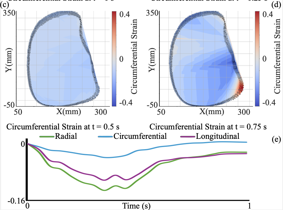
Abstract: Cardiovascular deaths are projected to increase from approximately 20.5 million in 2025 to 35.6 million by 2050 if current trends in risk factors continue. Early detection is critical for effective treatment and management. While conventional metrics such as ejection fraction provide useful information on cardiac function, they often lack the sensitivity needed to detect early-stage abnormalities. Advances in medical image analysis now allow for more detailed assessment of myocardial strain, a promising marker for early dysfunction. In this study, we evaluated three computational approaches, including optical flow, deep learning, and finite element modeling (FEM), to estimate myocardial strain from 4D CT images. Our goal was to track myocardial motion and deformation throughout the cardiac cycle and compare the performance and advantages of each method. First, we implemented a 3D Lucas-Kanade optical flow algorithm to estimate frame-to-frame motion. Next, a deep learning model was employed to automatically identify and track heart wall movements. Finally, FEM was used to model biomechanical behavior and extract strain in radial, circumferential, and longitudinal directions. Each method demonstrated distinct strengths: optical flow offered speed and simplicity, deep learning handled complex motion patterns, and FEM provided high biomechanical fidelity. By comparing these techniques, our framework offers deeper insight into cardiac mechanics and supports the development of more personalized, non-invasive assessments of cardiac function for clinical use.
Publication Thumbnail

Monfared, M., Parks, K., Hollingsworth, L., Hung, J., Knight, J., Gamage, P.T., Taebi, A (2025).
Computational Analysis of Cerebral Aneurysm Flow Dynamics at the M2 Bifurcation Using the Low-Reynolds K-ω Turbulence Model.
ASME International Mechanical Engineering Conference and Exposition, Memphis, TN.
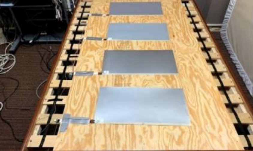
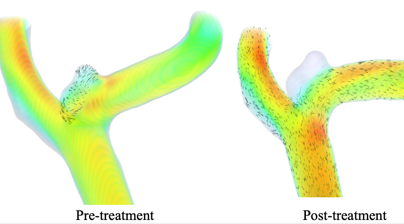
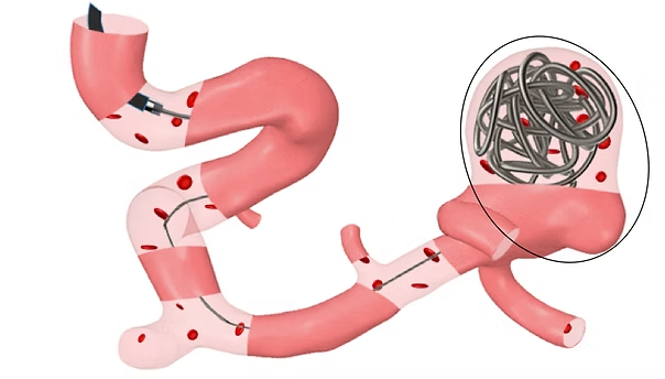
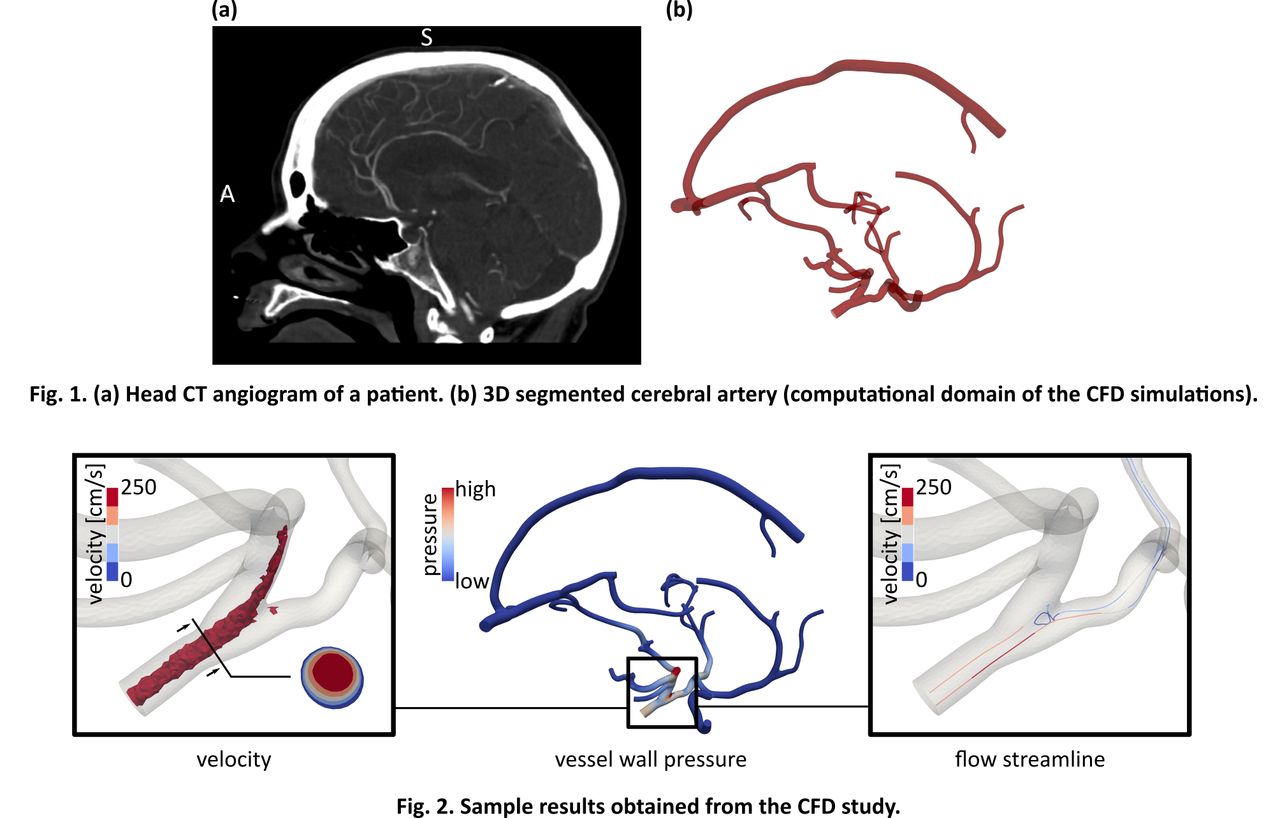
Abstract: Middle cerebral artery (MCA) aneurysms represent a significant clinical concern, accounting for up to 43% of all cerebral aneurysms and carrying a high risk of rupture, which may result in subarachnoid hemorrhage, permanent neurological damage, or death. More than 60% of MCA aneurysms occur at the M2 bifurcation, making it a critical site for hemodynamic analysis and treatment optimization. In this study, we investigate blood flow behavior in five patient-specific MCA aneurysm models using computational fluid dynamics (CFD). A porous medium approach is employed to simulate endovascular coiling, and physiologically realistic outlet boundary conditions are applied via the three-element Windkessel model. To account for transitional and turbulent flow features common at bifurcations and within aneurysm sacs, the low-Reynolds k–ω SST turbulence model is utilized. Mesh independence is verified following ASME guidelines, using three levels of refinement and evaluating the grid convergence index to ensure the accuracy of velocity and pressure predictions. Simulations before and after coiling show a marked reduction in intra-aneurysmal velocity and pressure, alongside a consistent decrease in wall shear stress near the aneurysm neck. These hemodynamic changes indicate effective isolation of the aneurysm sac from high flow regions, potentially reducing rupture risk. The results demonstrate the capability of CFD-based hemodynamic assessment for treatment planning of coil embolization. This framework supports more accurate and patient-specific evaluation of treatment strategies for cerebral aneurysms.
Publication Thumbnail

Esfahani, R.T., Loghmani, A, Akhavan, A., Taebi, A. (2025).
Evaluation of Signal Processing and Deep Learning Methods for Inter-Beat Interval Extraction from Ballistocardiography Signals.
Journal of Computational Methods in Engineering, 44(2): 49-61.​

Abstract: Cardiovascular diseases remain the leading cause of mortality worldwide, highlighting the critical need for continuous and non-invasive monitoring of cardiac function to enable early detection and effective management. Ballistocardiography (BCG), which captures the mechanical forces associated with cardiac activity, holds great promise for unobtrusive heart monitoring in daily-life settings without requiring direct electrode contact. However, the inherent complexity and high susceptibility to noise in BCG signals make the accurate extraction of key cardiac parameters—particularly inter-beat intervals (IBIs)—a challenging task. This study presents a comprehensive evaluation of five distinct signal processing and deep learning approaches for IBI estimation from BCG signals, validated against synchronized electrocardiogram (ECG) recordings. In contrast to the previous works, we employ a publicly available dataset distinct from those commonly used, enabling a broader assessment of method generalizability—particularly for the CLIE algorithm. The evaluated methods include: Continuous Local Interval Estimator (CLIE), CLIE with adaptive windowing, Multilayer Perceptron (MLP), Convolutional Neural Network (CNN), and Bidirectional Long Short-Term Memory (BiLSTM) network. For the deep learning methods (MLP and CNN), we propose novel network architectures specifically tailored to the characteristics of BCG signals, leading to improved performance compared to conventional designs. Furthermore, our BiLSTM-based method not only incorporates testing on a dataset different from that of previous reference studies, but also focuses on the accurate prediction of R-peak locations in the BCG signal, from which IBIs are subsequently derived. Evaluation based on Mean Absolute Error (MAE), 95th percentile error, and correlation coefficient shows that the CLIE method achieved the best overall IBI estimation accuracy, with an MAE of 28.7 milliseconds and the highest correlation coefficient (0.77). The BiLSTM method, while having a slightly higher MAE (40.1 milliseconds), demonstrated superior robustness to outliers by achieving the lowest 95th percentile error (9.5%). The MLP and CNN methods showed moderate performance, and the adaptive windowing variant of CLIE performed the worst. These findings demonstrate that accurate IBI extraction from BCG signals is feasible, and that both the CLIE and BiLSTM approaches are promising candidates for implementation in intelligent, home-based cardiac monitoring systems—offering, respectively, high accuracy and strong resilience to large errors.
Publication Thumbnail

Taebi, A., Rahman, M.M. (2025).
MSCardio seismocardiography dataset: Initial insights from remote monitoring of cardiovascular-induced chest vibrations via smartphones.
Data in Brief, 61:111889.​

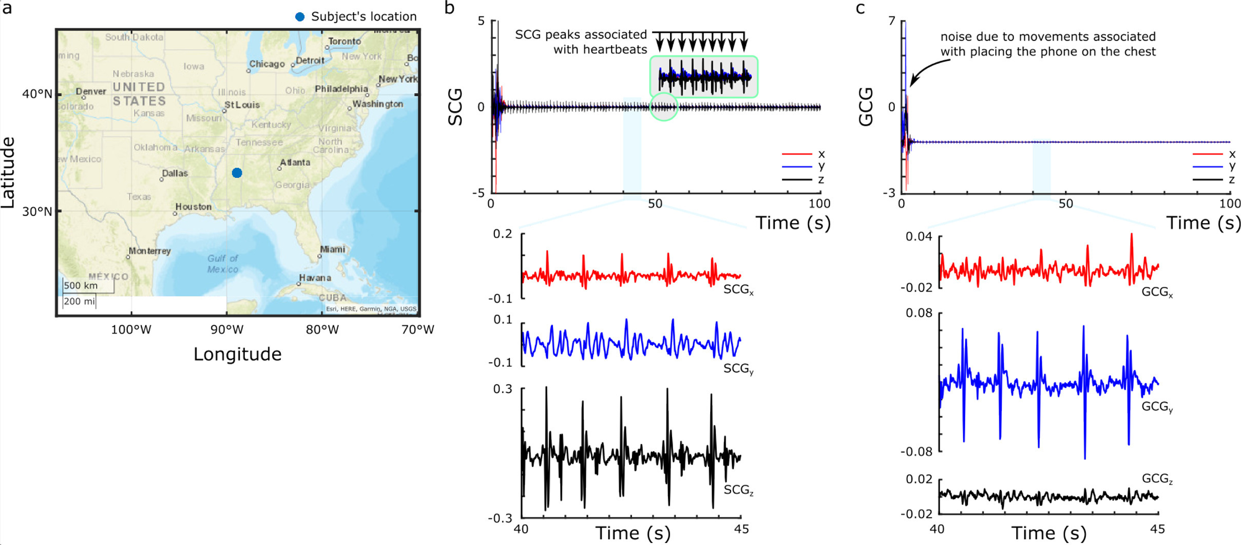
Abstract: The Mississippi State Remote Cardiovascular Monitoring (MSCardio) Study leverages smartphones to remotely collect seismocardiography (SCG) and gyrocardiography data from participants. The study employs an app that uses embedded sensors to record multi-dimensional vibrations, along with self-reported lifestyle and health data. Metadata is also captured to provide contextual information for analysis. This paper outlines the progress made during the first 250 days of the study, including the methodology, data collection process, and preliminary findings. Additionally, the SCG dataset collected during this period is publicly shared to support further research. These results highlight the potential of smartphone-based systems for scalable and accessible cardiovascular health monitoring.
Publication Thumbnail

Rahman, M., Taebi, A. (2025).
Contactless heart rate and heart rate variability estimation from neck videos.
47th Annual International Conference of the IEEE Engineering in Medicine and Biology Society, Copenhagen, Denmark.
Abstract: Video-based pulse extraction is a non-contact technique for estimating physiological signals from video recordings. While traditional approaches focus on facial regions due to their high vascularity and accessibility, this study explored the feasibility of extracting pulse signals from neck region, an area less studied in the literature. Nech and chest video data were collected from 14 healthy subjects during breath-hold. A region of interest on the neck was tracked using a template-based algorithm, and pixel intensity variations within this region were processed using six established video-based pulse extraction methods: GREEN, the chrominance-based method, plant-orthogonal-to-skin method, orthogonal matrix image transformation method, independent component analysis method, and local group invariance method. The extracted pulse signals were validated against synchronized electrocardiogram (ECG) recordings. Among the methods, GREEN exhibited the highest agreement with ECG-derived heart rate (HR), with a bias of -4.89 bpm and limits of agreement (LoA) ranging from 29.97 to 20.17 bpm. After excluding two subjects with significant noise interference, the agreement improved to a bias of -1.42 bpm and LoA of -4.95 to 2.09 bpm. HR variability (HRV) was assessed using SDNN. SDNN values from GREEN method were generally higher than those from ECG, likely due to signal differences, motion artifacts, and the short duration (1015 seconds) of the recordings, which may have inflated variability estimates. Findings suggested that the neck region, particularly when using the GREEN method, is a viable site for video-based pulse signal extraction and HR estimation. However, variations in SDNN underscore the need for further refinement in video-based pulse extraction techniques. Expanding this technology to the neck region offers promising applications in remote health monitoring and physiological assessment, particularly to mitigate the privacy concerns of facial recordings.
Publication Thumbnail

Ahmed, M.H., Panchookian, J., Grillo, M., Weerasinghe, Y., Taebi, A., Qadri, F., Gamage, P., Kaya, M. (2025).
Stress Classification Through Simultaneous EEG, Heart Rate Variability, and EMG Monitoring.
IEEE Medical Measurements & Applications, Chania, Greece.
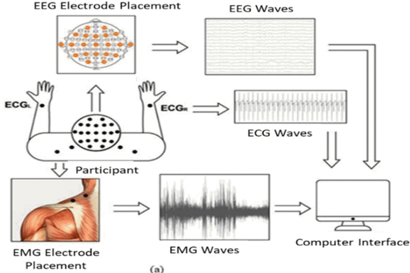
Abstract: Stress has significant effects on health, yet there is limited research on effective methods for quantifying stress detection. Monitoring physiological changes presents a promising approach to stress management. This study compares the effectiveness of electroencephalography (EEG), electrocardiography (ECG)-derived heart rate variability (HRV), and trapezius muscle electromyography (EMG) in stress classification. Sixteen healthy participants (ages 18–46) completed three sessions in a controlled environment. Baseline activity was compared to stress-induced changes during a Stroop color word test and mental arithmetic task. EEG, HRV, and EMG features were analyzed in 30-second intervals to assess their ability to detect stress. EEG features were found to be the most effective, followed by HRV and EMG. Machine learning techniques (SVM, KNN, neural network, and random forest) were applied for subject-specific classification. EEG achieved the highest accuracy (86.45 ± 7.22%), while HRV and EMG yielded similar accuracies (77.36 ± 9.10% and 81.84 ± 6.13%, respectively). When combining HRV and EMG features, an accuracy of 87.51 ± 7.18% was achieved, comparable to EEG. These findings suggest that wearable sensors utilizing EMG and HRV could effectively detect stress without the need for EEG. This approach could open up new avenues for stress management in real-world settings. Future studies with larger sample sizes will work towards developing a universal stress classification model.
Publication Thumbnail

Monfared, M., Gamage, P.T., Loghmani, A., Taebi, A. (2025).
Computational Modeling of Cardiovascular‐Induced Chest Vibrations: A Review and Practical Guide for Seismocardiography Simulation.
International Journal for Numerical Methods in Biomedical Engineering, 41(5): e70047.​

Abstract: This paper presents a comprehensive examination of finite element modeling (FEM) approaches for seismocardiography (SCG), a non-invasive method for assessing cardiac function through chest surface vibrations. The paper provides a comparative analysis of existing FEM approaches, exploring the strengths and challenges of various modeling choices in the literature. Additionally, we introduce a sample framework for developing FEM models of SCG, detailing key methodologies from governing equations and meshing techniques to boundary conditions and material property selection. This framework serves as a guide for researchers aiming to create accurate models of SCG signal propagation and offers insights into capturing complex cardiac mechanics and their transmission to the chest surface. By consolidating the current methodologies, this paper aims to establish a reference point for advancing FEM-based SCG modeling, ultimately improving our understanding of SCG waveforms and enhancing their reliability and applicability in cardiovascular health assessment.
Publication Thumbnail

Rahman, M.M., Kakavand, B., Wurm, W., Holman, W., Movahed, M.R., Taebi, A. (2025).
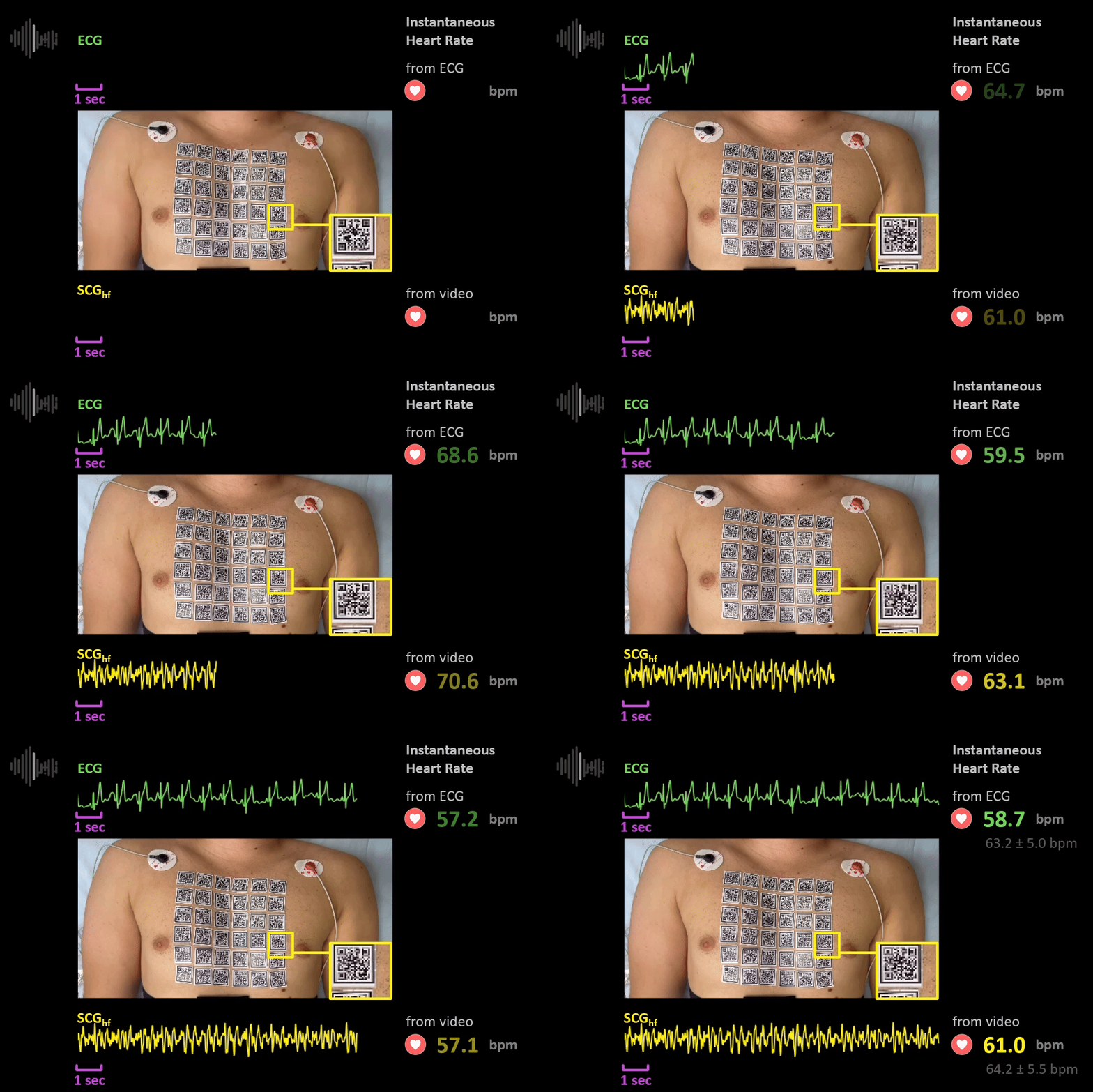
From video to vital signs: a new method for contactless multichannel seismocardiography.
npj Cardiovascular Health, 2, 1.
Abstract: Seismocardiography (SCG) is a technique that non-invasively measures the chest wall’s local vibrations caused by the heart’s mechanical activity. Traditionally, SCG signals have been recorded using accelerometers placed at a single location on the chest wall. This study presents an innovative, cost-effective SCG method that utilizes standard smartphone videos to capture data from multiple chest locations. The analysis of vibrations from multiple points can offer a more thorough understanding of the heart’s mechanical activity compared to signals obtained solely from a single chest location. Our approach employs computer vision and deep learning techniques to extract and improve the resolution of multichannel SCG maps obtained by video capture of chest movement. We attached a grid of patterned stickers to the chest surface and recorded videos of chest movements during different respiratory phases. Using a deep learning-based object detector and a template tracking method, we tracked the stickers across video frames and extracted the corresponding SCG signals from sticker displacements. We also developed a robust algorithm to estimate heart rate (HR) from these chest videos and identify the optimal chest location for HR estimation. The method was tested on 28 chest videos captured from 14 healthy participants. The results demonstrated that our method effectively extracted multichannel SCG maps and enhanced their resolution with a mean squared error of 0.1078 and 0.0418 for right-to-left and head-to-foot SCG signals, respectively. We observed intersubject chest vibration patterns corresponding to cardiac events including opening and closure of the heart valves. Moreover, our algorithm accurately estimated HR from 1968 SCG signals extracted from the videos compared to the gold-standard HR measured from each subject’s electrocardiogram (bias ± 1.96 SD = 0.04 ± 2.14 bpm; r = 0.99, p < 0.001). The findings from this study underscore the potential of our approach in developing a cardiac monitoring tool using a smartphone that would be widely accessible to the general public and might provide more timely detection of diseases.
Publication Thumbnail

Monfared, M., Hollingsworth, L., Gamage, P.T., Taebi, A. (2025).
Assessing Hemodynamic Changes in Cerebral Aneurysms Post Coil Embolization: A Preliminary Investigation.
ASME Journal of Engineering and Science in Medical Diagnostics and Therapy, 8(3): 031014.​

Abstract: A cerebral aneurysm is a weakened area in the wall of a blood vessel within the brain that causes the vessel to bulge outwards. The choice of treatment depends on various factors including the size and location of the aneurysm and the risk of rupture, and should be tailored to individual situations rather than following a universal approach due to their variability among individuals. This approach provides the possibility of choosing precise treatment plans that are proper for each patient’s circumstances. The utilization of computational fluid dynamics (CFD) allows for the prediction of after-surgery hemodynamic changes before any surgical approach. The goal of this research was to investigate the flow characteristics and hemodynamic parameters in cerebral arteries before and after endovascular embolization treatments using CFD in three patient-specific geometries with at least one aneurysm at the middle cerebral artery’s bifurcation. To model the coiling, the computational domain was divided into two fluid domains, a general fluid domain consisting of the parent arteries and a porous domain inside the aneurysm. CFD modeling was conducted to simulate blood flow in pre- and postcoiling scenarios. Results showed that the coiling model with different porosity values led to a redirection of blood flow away from the aneurysm sac. Additionally, changes in shear stress indicated potential alterations in susceptibility to vascular remodeling. In conclusion, these findings indicated that utilizing CFD modeling for simulating blood flow in patient-specific cerebral arteries could be utilized to predict hemodynamic parameters before any treatment.
Publication Thumbnail

Rahman, M., Taebi, A. (2024).
Extracting Cardiovascular-Induced Chest Vibrations from Ordinary Chest Videos: A Comparative Study.
IEEE Signal Processing in Medicine and Biology.
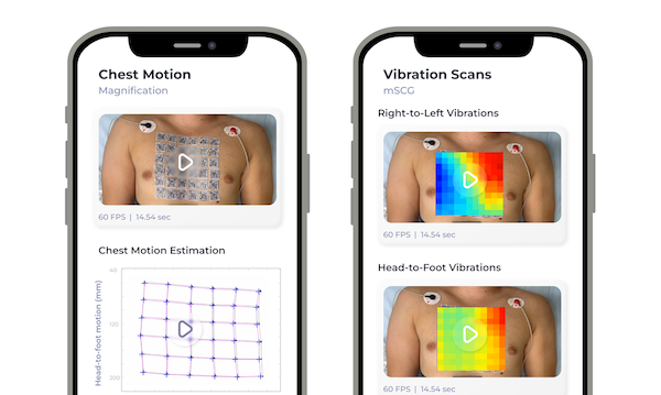
Abstract: Seismocardiography (SCG) has attracted significant interest for monitoring cardiac health and diagnosing cardiovascular conditions. While traditional SCG methods rely on uncomfortable chest-mounted accelerometers, recent research explores non-contact approaches, including analyzing video recordings of the chest. In this study, three computer vision-based methods including Lucas-Kanade optical flow, template tracking, and Gunnar-Farneback optical flow were evaluated for extracting SCG signals from ordinary camera-recorded chest videos. The study focused on right-to-left and head-to-foot SCG signals obtained from 13 healthy subjects during breath-hold at the end of exhalation and inhalation. Comparative analysis was performed by calculating the mean squared error (MSE) and root MSE (RMSE) between the vision-based SCG signals and the gold-standard accelerometer signals. Visual and quantitative analyses showed that the Lucas-Kanade and template tracking methods estimated vision-based SCG signals closely resembling the accelerometer data, particularly in the head-to-foot direction. The Lucas-Kanade method had MSE values ranging from 0.14 to 0.93, RMSE values from 0.38 to 0.96, average correlation values of 0.82±0.09. The template tracking method showed MSE values between 0.12 to 0.94, RMSE values from 0.35 to 0.97, and average correlation values of 0.83±0.10. In comparison, the Farneback method had higher MSE values ranging from 0.20 to 1.07, RMSE values from 0.44 to 1.03, and average correlation values of 0.76±0.11. These results suggest the effectiveness of Lucas-Kanade and template tracking methods for non-contact SCG signal extraction from chest video data.
Publication Thumbnail

Ruckman, S., Taghva, F., Taebi, A. (2024).
Parent-Centered Design: A Preliminary Usability Study on Newborn Home Cardiac Monitoring.
2024 IEEE Signal Processing in Medicine and Biology.
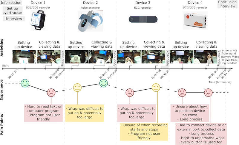
Abstract: Current measurement techniques in the medical field, such as electrocardiography (ECG) and pulse oximetry, are critical for monitoring the cardiovascular health of newborns. Although there are devices on the market capable of measuring these signals, they are often not designed for use with newborns. This study conducted a preliminary usability test of four cardiovascular monitors for use with a newborn model. The findings indicated that users prefer a simple, wireless device that can be securely attached to a child to minimize movement. Additionally, they value a guided process and straightforward, user-friendly software. Understanding user preferences can facilitate developing an affordable, at-home monitoring device tailored for newborns which in turn may enhance early detection and management of congenital heart diseases (CHDs).
Publication Thumbnail

Mann, A., Rahman, M.M., Vanga, V., Gamage, P.T., Taebi, A. (2024).
Variation of Seismocardiogram-Derived Cardiac Time Intervals and Heart Rate Variability Metrics Across the Sternum.
ASME J of Medical Devices, 18(4): 044502.
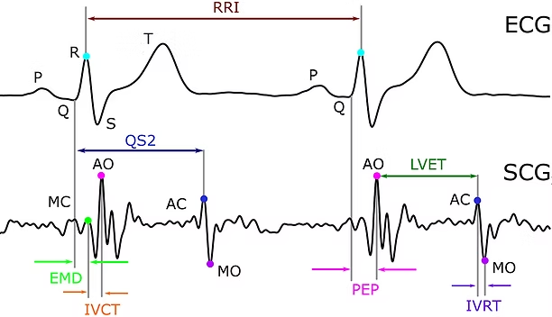
Abstract: Cardiac time intervals (CTIs) are vital indicators of cardiac health and can be noninvasively assessed using a combination of electrocardiography (ECG) and seismocardiography (SCG), a method of capturing cardiac-induced chest vibrations via accelerometers. SCG signals can be measured from different chest locations. However, more investigations are needed to evaluate the impact of sensor placement on SCG-derived cardiac parameters. This study investigates the effect of accelerometer placement along the sternum on SCG-derived CTI estimations and heart rate variability (HRV) parameters. A semi-automated algorithm was developed to detect SCG fiducial points and seven CTIs from thirteen healthy individuals. Comparative analysis with manually selected peaks and gold-standard ECG was conducted to assess fiducial point detection accuracy. Results indicate the highest recall and precision in aortic valve opening (0.84–1.00 and 0.96–1.00, respectively) and mitral valve closure (0.77–1.00 and 0.93–1.00, respectively) detection. Aortic valve closure (0.43–1.00 and 0.61–1.00, respectively) and mitral valve opening (0.64–1.00 and 0.91–1.00, respectively) detection, although slightly less accurate due to signal intensity variations, demonstrated overall effectiveness compared to manually selected peaks. Furthermore, SCG-derived heart rates showed a high correlation coefficient (r > 0.9) with the gold-standard ECG heart rates. Single-factor ANOVA revealed significant differences (p < 0.05) in SCG-derived CTI estimations based on sensor locations on the sternum, highlighting the importance of sensor placement for accurate assessments.
Publication Thumbnail

Monfared, M., Gamage, P.T., Taebi, A. (2024).
Investigating Seismocardiogram Patterns: A Computational Modeling of Cardiac Wall Motion Propagation to the Chest Surface
ASME International Mechanical Engineering Conference and Exposition, Portland, OR, V004T06A006.

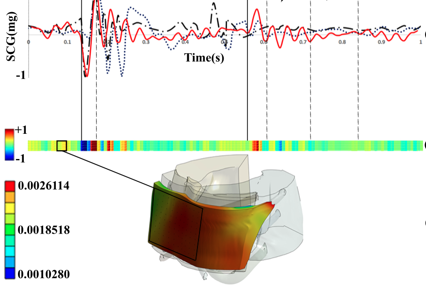
Abstract: Cardiovascular diseases, the leading cause of global mortality, demand refined diagnostic methods. Seismocardiography (SCG), a noninvasive method of measuring cardiovascular-induced vibrations on the chest surface, offers promise in assessing cardiac function. The cardiac wall movements are transmitted to the organs around the heart and eventually damped onto the chest surface, where they manifest as visible vibrations. These chest surface vibrations can be measured using an accelerometer via SCG. Although SCG signals are widely used in literature, further investigations are needed to understand the genesis of their patterns under different pathophysiological conditions. The goal of this study is to improve our understanding of the origin of SCG signals by simulating the transmission of cardiac motion reaching the chest surface using finite element method, and linking back the patterns of the simulated SCG signals to specific cardiac events. The computational domain, extracted from 4D computed tomography (CT) images of a healthy subject, comprised the lungs, ribcage, and chest muscles and fat. Using the Lukas-Kanade algorithm, the cardiac wall motion was extracted from the 4D CT scan images and was used as a displacement boundary condition. The elastic material properties were assigned to the lungs, muscles, fat, and rib cage. The dorsoventral SCG component from the finite element modeling was compared with two actual SCG signals obtained from the literature. The left ventricular volume was also calculated from the CT scans and was used to interpret the SCG waveforms. Important cardiac phases were labeled on the SCG signal extracted from the computationally modeled acceleration map near the xiphoid. This type of analysis can provide insights into various cardiac parameters and SCG patterns corresponding to the mitral valve closing, mitral valve opening, aortic valve opening, and aortic valve closure. These findings suggested the effectiveness of this modeling approach in understanding the underlying sources of the SCG waveforms.
Publication Thumbnail

Rahman, M., Taghva, F., Taebi, A. (2024).
Novel Contactless and AI-Based Method Can Determine Heart Rate and Cardiac-Induced Vibrations of Chest.
Circulation 150 (Suppl_1): A4112524.

Publication Thumbnail

Taebi, A. (2024).
Computational Fluid Dynamics in Medicine and Biology.
Bioengineering 11 (11), 1168.

Editor's Choice
Publication Thumbnail

Ahmed, M., Weerasinghe, Y., Grillo, M., Taebi, A., Kaya, M., Gamage, P.T. (2024).
Feasibility of Left Ventricular Function Assessment via Precordial Vibrations in Heart Failure Patients.
ASME International Mechanical Engineering Conference and Exposition, Portland, OR, V004T06A007.
Abstract: Heart failure (HF) is a prevalent and potentially life-threatening condition characterized by the heart’s inability to pump blood effectively. Managing HF requires careful monitoring of left ventricular function (LVF), necessitating timely interventions to prevent adverse outcomes. Traditional methods of monitoring LVF can be invasive, costly, and limited to clinical settings which poses challenges for continuous monitoring and early detection of HF progression. This study aims to evaluate the feasibility of estimating left ventricular ejection fraction (LVEF) from precordial vibration signals. Precordial vibration data from 70 patients with varying levels of HF were analyzed. Seismocardiography (SCG) signals were processed to extract features and train machine-learning regression models. The predicted LVEF values from the models were compared with LVEF data obtained from echocardiography, the gold standard for LVF assessment. Among trained machine learning models, Gaussian process regression indicated the greatest correlation between the estimated and obtained values with the least mean squared error. Variations in the precordial vibrations were observed to correlate with the severity of valvular dysfunction, highlighting the sensitivity of this approach. In conclusion, precordial vibration signals have the potential to offer valuable insights into both myocardial and valvular function in HF patients. Precordial vibration analysis shows promise as a non-invasive, cost-effective method for continuous monitoring of LVF in HF patients. Further research and validation are warranted to explore its clinical applicability and potential integration into routine HF management protocols, with the ultimate goal of improving patient outcomes and quality of life.
Publication Thumbnail

Weerasinghe, Y., Sheppard, C., Ahmed, M., Grillo, M., Taebi, A., Wilder, D., Kaya, M., Gamage, P.T. (2024).
Markerless Pose Estimation and Wearable Sensor Technology for Enhanced Diagnosis and Monitoring of Idiopathic Toe Walking.
ASME International Mechanical Engineering Conference and Exposition, Portland, OR, V004T06A019.
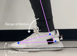
Abstract: Idiopathic Toe Walking (ITW) is a common condition characterized by an individual’s preference for walking on their toes without any identifiable medical reasons. Children usually outgrow this habit; however, those who don’t are often affected by certain conditions such as cerebral palsy, muscular dystrophy, and autism spectrum disorder. If not addressed, ITW can lead to physiological complications such as skeletal and muscular deformations. Identifying and monitoring toe walking patterns is only accessible through qualitative observational gait analysis and electromyography (EMG). This approach’s main objective is to provide quantitative means to analyze ITW and diagnose and monitor patient rehabilitation. The markerless pose estimation approach analyzes the ankle’s range of motion to identify normal and abnormal gait patterns. In addition to the markerless pose estimation, a 6-axis accelerometer and gyroscope inertial measurement unit (IMU) allowed for the continuous monitoring of motion values, which constitute the identification of idiopathic toe walking. Both noninvasive approaches provided quantifiable angles, allowing for convenient and continuous ITW monitoring. By employing markerless pose estimation coupled with a wearable sensor, ITW could be monitored, enhancing the accessibility of continuous monitoring for gait irregularities.
Publication Thumbnail

Rahman, M., Taebi, A. (2024).
Contactless seismocardiography via Gunnar-Farneback optical flow.
IEEE 20th International Conference on Body Sensor Networks, Chicago, IL.
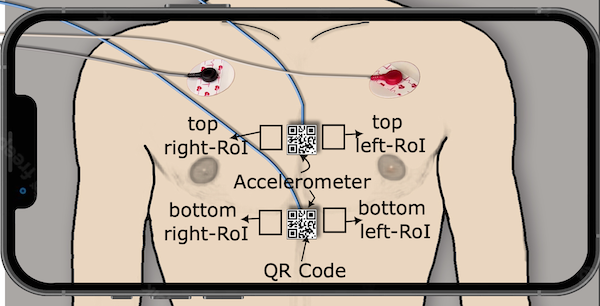
Abstract: Seismocardiography (SCG) has gained significant attention due to its potential applications in monitoring cardiac health and diagnosing cardiovascular conditions. Conventional SCG methods rely on accelerometers attached to the chest, which can be uncomfortable or inconvenient. In recent years, researchers have explored non-contact methods to capture SCG signals, and one promising approach involves analyzing video recordings of the chest. In this study, we investigate a vision-based method based on the Gunnar-Farneback optical flow to extract SCG signals from the chest skin movements recorded by a smartphone camera. We compared the SCG signals extracted from the chest videos of four healthy subjects with those obtained from accelerometers and our previous method based on sticker tracking. Our results demonstrated that the vision-based SCG signals extracted by the proposed method closely resembled those from accelerometers and stickers, although these signals were captured from slightly different locations. The mean squared error between the vision-based SCG signals and accelerometer-based signals was found to be within a reasonable range, especially between signals on head-to-foot direction (0.2<MSE<1.5). Additionally, heart rates derived from the vision-based SCG exhibited good agreement with the gold-standard ECG measurements, with a mean difference of 0.8 bpm. These results indicate the potential of this non-invasive method in health monitoring and diagnostics.
Publication Thumbnail

Rahman, M., Mann, A., Taebi, A. (2024).
ECG-Free Assessment of Cardiac Valve Events Using Seismocardiography.
IEEE 20th International Conference on Body Sensor Networks, Chicago, IL.
Abstract: Seismocardiogram (SCG) signals can play a crucial role in remote cardiac monitoring, capturing important events such as aortic valve opening (AO) and mitral valve closure (MC). However, existing SCG methods for detecting AO and MC typically rely on electrocardiogram (ECG) data. In this study, we propose an innovative approach to identify AO and MC events in SCG signals without the need for ECG information. Our method utilized a template bank, which consists of signal templates extracted from SCG waveforms of 5 healthy subjects. These templates represent characteristic features of a heart cycle. When analyzing new, unseen SCG signals from another group of 6 healthy subjects, we employ these templates to accurately detect cardiac cycles and subsequently pinpoint AO and MC events. Our results demonstrate the effectiveness of the proposed template bank approach in achieving ECG-independent AO and MC detection, laying the groundwork for more convenient remote cardiovascular assessment.
Publication Thumbnail

Ahmed, M.H., Grillo, M., Taebi, A., Kaya, M., Gamage, P.T. (2024).
A Comprehensive Analysis of Trapezius Muscle EMG Activity in Relation to Stress and Meditation.
BioMedInformatics, 4(2): 1047-1058.
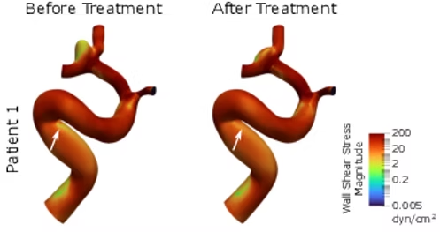
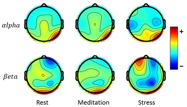
Abstract: Introduction: This study analyzes the efficacy of trapezius muscle electromyography (EMG) in discerning mental states, namely stress and meditation. Methods: Fifteen healthy participants were monitored to assess their physiological responses to mental stressors and meditation. Sensors were affixed to both the right and left trapezius muscles to capture EMG signals, while simultaneous electroencephalography (EEG) was conducted to validate cognitive states. Results: Our analysis of various EMG features, considering frequency ranges and sensor positioning, revealed significant changes in trapezius muscle activity during stress and meditation. Notably, low-frequency EMG features facilitated enhanced stress detection. For accurate stress identification, sensor configurations can be limited to the right trapezius muscle. Furthermore, the introduction of a novel method for determining asymmetry in EMG features suggests that applying sensors on bilateral trapezius muscles can improve the detection of mental states. Conclusion: This research presents a promising avenue for efficient cognitive state monitoring through compact and convenient sensing.
Publication Thumbnail

Davidson, H., Scardino, B., Gamage, P.T., Taebi, A. (2024).
Variations of Middle Cerebral Artery Hemodynamics Due to Aneurysm Clipping Surgery.
ASME Journal of Engineering and Science in Medical Diagnostics and Therapy, 7(1): 011008.
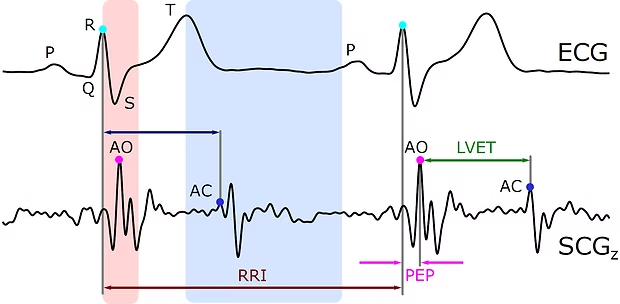
Abstract: Cerebral aneurysms are potentially life-threatening cerebrovascular conditions where a weakened blood vessel in the brain bulges or protrudes over time. The most common way to treat aneurysms is surgical clipping, an approach where blood flow to the aneurysm is blocked by a permanently placed clip on the artery. However, not all aneurysms are identical; thus, there has been a need for patient-specific treatment options, where each aneurysm is treated based on its individual characteristics. Computational fluid dynamics (CFD) modeling can offer insights to predict how different treatment procedures will affect cerebral hemodynamics. In that regard, the goal of this pilot study was to investigate the flow characteristics and hemodynamic parameters in cerebral arteries before and after neurosurgical clipping. For this purpose, two patient-specific cerebral artery geometries with at least one aneurysm at the middle cerebral artery bifurcation were selected from an online dataset. A companion postclipping model was created for each geometry by removing the aneurysm from the original geometry. Tetrahedral mesh elements were then generated and CFD simulations were conducted to compare the blood velocity profile, secondary flow, flow streamline, and wall shear stress in the computational models with and without aneurysm. Results showed that the clipping treatment led to changes in the velocity profiles, secondary flow structures, and wall shear stress in the middle cerebral artery. In conclusion, our results suggest that CFD modeling can assist in predicting hemodynamic parameters prior to treatment, thus facilitating more tailored planning for each patient’s treatment.
Publication Thumbnail

Mann, A., Gamage, P.T., Kakavand, B., Taebi, A. (2024).
Exploring the Impact of Sensor Location On Seismocardiography-Derived Cardiac Time Intervals.
ASME Journal of Engineering and Science in Medical Diagnostics and Therapy, 7(1): 011007.
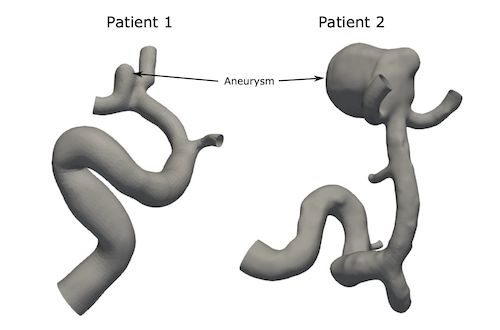
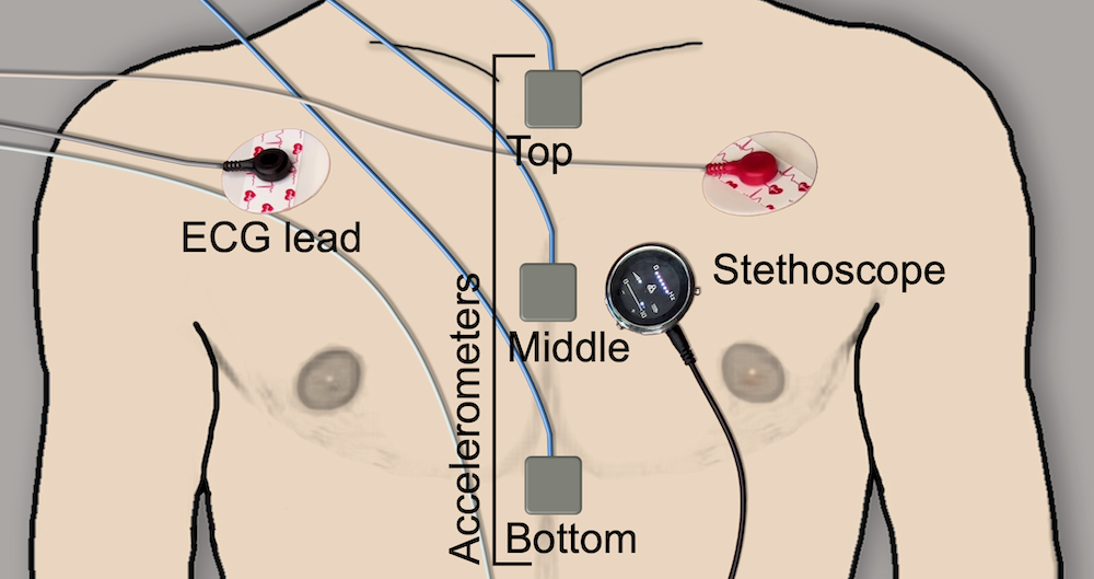
Abstract: Cardiac time intervals (CTIs) are important parameters for evaluating cardiac function and can be measured noninvasively through electrocardiography (ECG) and seismocardiography (SCG). SCG signals exhibit distinct spectrotemporal characteristics when acquired from various locations on the chest. Thus, this study aimed to explore how SCG measurement location affects the estimation of SCG-based CTIs. ECG and SCG signals were acquired from 14 healthy adults, with three accelerometers placed on the top, middle, and bottom of the sternum. A custom-built algorithm was developed to estimate heart rates (HRs) from ECG (HRECG) and SCG (HRSCG) signals. Moreover, SCG fiducial points and CTIs, including aortic valve opening and closure, R-R interval, pre-ejection period, left ventricular ejection time, and electromechanical systole, were estimated from the SCG signals at different sternal locations. The average and correlation coefficient (R2) of the CTIs and HRs derived from all three locations were compared, along with the analysis of mean differences for the CTIs and their corresponding sensor locations. The results indicated strong correlations between HRECG and HRSCG, with average R2 values of 0.9930, 0.9968, and 0.9790 for the top, middle, and bottom sternal locations, respectively. Additionally, the study demonstrated that SCG-based CTIs varied depending on the SCG measurement locations. In conclusion, these findings underscore the importance of establishing consistent protocols for reporting CTIs based on SCG. Furthermore, they call for further investigation to compare estimated CTIs with gold-standard methods like echocardiography to identify the best SCG measurement location for accurate CTI estimations.
Publication Thumbnail

Davidson, H., Scardino, B., Hollingsworth, L., Gamage, P.T., Taebi, A. (2023).
A Comparative Study of Middle Cerebral Artery Hemodynamics Pre- and Post-Clipping of Cerebral Aneurysm.
ASME International Mechanical Engineering Conference and Exposition, New Orleans, LA, V005T06A069.
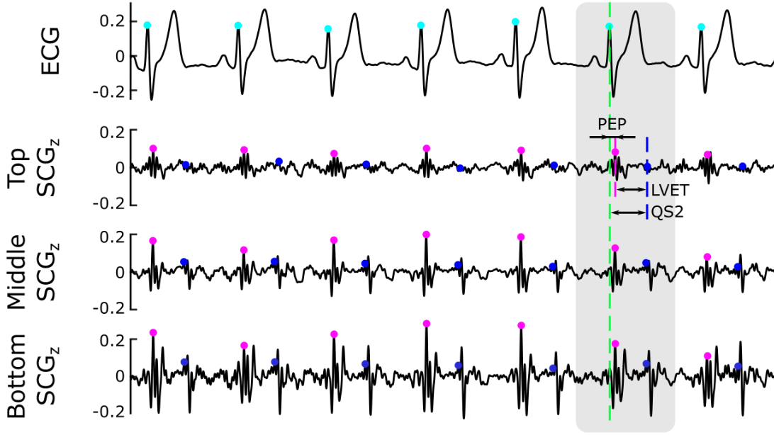
Abstract: Cerebral aneurysms (CAs), also referred to as brain or intracranial aneurysms, are potentially life-threatening cerebrovascular conditions where a weakened blood vessel in the brain bulges or protrudes over time. CAs pose a serious health concern if left untreated, as they can lead to rupture, permanent neurological deficit, and fatal aneurysmal subarachnoid hemorrhage. Currently, the most common way to treat aneurysms is surgical clipping, an approach where blood flow to the aneurysm is blocked by a permanently placed clip on the artery. However, not all aneurysms are identical; thus, there has been a need for patient-specific treatment options, a method where each aneurysm is treated based on its individual characteristics. This method allows for a more accurate treatment plan that can be tailored for each patient. In that regard, computational fluid dynamics (CFD) modeling can offer insights to predict how different treatment procedures will affect cerebral hemodynamics. More specifically, during treatment planning, patient-specific CFD modeling of blood flow can aid in further understanding the pathophysiology of CAs and assess aneurismal flow in a range of scenarios and physiological conditions. The goal of this pilot study was to investigate the flow characteristics and hemodynamic parameters in cerebral arteries before and after neurosurgical clipping treatments. For this purpose, two patient-specific cerebral artery geometries were selected from a dataset created by the @neurIST project. Each geometry included at least one aneurysm at the middle cerebral artery bifurcation. Open-source software, ParaView and OpenFlipper, was used to construct and smooth the 3D models, i.e., computational models. A companion model was also created for each geometry by removing the aneurysm from the original geometry and was used as the post-clipping artery model. Tetrahedral mesh elements were then generated and CFD simulations were conducted in SimVascular. Finally, the simulation results were post-processed in ParaView to visualize and compare the blood velocity profile, secondary flow, flow streamline, and wall shear stress in the computational models with and without aneurysm (corresponding to pre- and post-clipping). Results showed that the clipping treatment led to changes in the velocity profiles, secondary flow structures, and wall shear stress in the middle cerebral artery. In conclusion, our results suggest that employing CFD modeling to simulate blood flow in patient-specific cerebral arteries can assist in predicting hemodynamic parameters prior to treatment, thus facilitating more tailored planning for each individual patient’s treatment. This, in turn, can potentially reduce the risks of serious complications and improve clinical outcomes. To further advance this research, future directions could involve exploring additional models of cerebral aneurysms, incorporating more realistic boundary conditions, and validating the current simulation results.
Publication Thumbnail

Mann, A., Kakavand, B., Gamage, P.T., Taebi, A. (2023).
Effect of Measurement Location on Cardiac Time Intervals Estimated by Seismocardiography.
ASME International Mechanical Engineering Conference and Exposition, New Orleans, LA, V005T06A070.
Abstract: Cardiac time intervals (CTIs) are important parameters for evaluating cardiac function and can be measured noninvasively through electrocardiography (ECG) and seismocardiography (SCG). SCG signals exhibit distinct spectrotemporal characteristics when acquired from various locations on the chest. Thus, this study aimed to explore how SCG measurement location affects the estimation of SCG-based CTIs. ECG and SCG signals were acquired from 14 healthy adults, with three accelerometers placed on the top, middle, and bottom of the sternum. A custom-built algorithm was developed to estimate heart rates (HRs) from ECG (HRECG) and SCG (HRSCG) signals. Moreover, SCG fiducial points and CTIs, including aortic valve opening and closure, R-R interval, pre-ejection period, left ventricular ejection time, and electromechanical systole, were estimated from the SCG signals at different sternal locations. The average and correlation coefficient (R2) of the CTIs and HRs derived from all three locations were compared, along with the analysis of mean differences for the CTIs and their corresponding sensor locations. The results indicated strong correlations between HRECG and HRSCG, with average R2 values of 0.9930, 0.9968, and 0.9790 for the top, middle, and bottom sternal locations, respectively. Additionally, the study demonstrated that SCG-based CTIs varied depending on the SCG measurement locations. In conclusion, these findings underscore the importance of establishing consistent protocols for reporting CTIs based on SCG. Furthermore, they call for further investigation to compare estimated CTIs with gold-standard methods like echocardiography to identify the best SCG measurement location for accurate CTI estimations.
best master’s paper award in the biomedical and biotechnology track
Publication Thumbnail

Ruckman, S., Bhatt, J., Cook, J., Gamage, P.T., Kakavand, B., Taebi, A. (2023).
Design, Prototype, and Evaluation of a Low-Cost Multimodal Device for Cardiovascular Monitoring.
ASME International Mechanical Engineering Conference and Exposition, New Orleans, LA, V005T06A022.
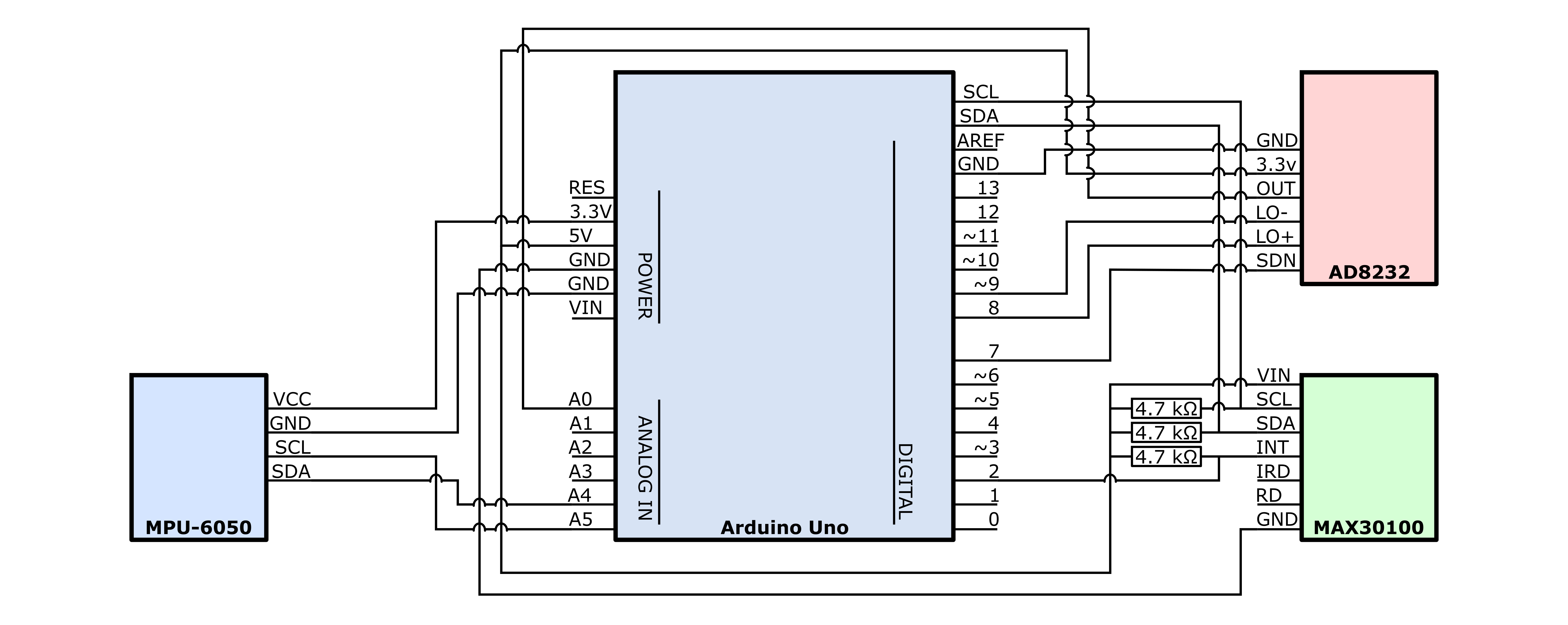
Abstract: Cardiovascular diseases (CVDs) are the leading cause of death in the United States. In most cases, these diseases go undiagnosed and untreated in the general population until the patient’s health is severely affected. Scanning for these diseases is generally done in a clinical setting, something that not everyone takes advantage of. These tests can also be expensive for many, especially in underserved areas. In that regard, the availability of a low-cost monitoring device can help in the early detection and management of cardiovascular diseases, leading to reduced morbidity, mortality, and associated healthcare costs. The goal of this pilot study is to construct and establish the feasibility of an inexpensive sensing device that is noninvasive and can be used to monitor cardiovascular functions outside of healthcare facilities. This device includes an MPU-6050 inertial measurement unit, an AD8232 signal conditioning block, a MAX30102 sensor, and a microcontroller to measure triaxial seismocardiogram (SCG), triaxial gyrocardiogram (GCG), electrocardiogram (ECG), and the oxygen level of the blood. The sensors were enclosed by a custom-designed case that is composed of various 3D printed parts. The combination of these different modalities assessed both electrical and mechanical aspects of cardiovascular activity. To evaluate the performance of the device, three adult subjects (both sexes: 2 males and 1 female; mean age: 26±5.29 years old) were recruited after institutional review board approval. The subjects were asked to lie in a supine position and breath normally during the experiment. The prototype was placed on the sternum around the 4th intercostal space of the subjects to acquire their SCG, GCG, and ECG signals, as well as the blood oxygen level. The ECG electrodes were placed under the right clavicle, left clavicle, and the lower right side of the abdomen. The signals of interest were also measured simultaneously using more sensitive, expensive, and commercially available sensors (as the gold standard). The signals collected from the prototype were then compared to the gold-standard signals using a similarity index based on dynamic time warping. Results demonstrated a high similarity between the two groups of signals, proving the feasibility of the proposed multimodal device. Future directions for this technology include wireless integration, increased compactness, reduced cost, and clinical testing on a larger and more diverse population including patients with cardiovascular issues. This device will ultimately provide an inexpensive and noninvasive method of scanning for CVDs in the general population.
Publication Thumbnail

Villa, A., Ahmad, M., Taebi, A., Dong, P., Gu, L., Gamage, P.T. (2023).
A Reduced Order Model for Estimation of Fractional Flow Reserve (FFR) in Coronary Artery Disease: Assessing the Impact of Side Branches.
ASME International Mechanical Engineering Conference and Exposition, New Orleans, LA, V005T06A046.
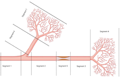
Abstract: Millions of people worldwide are affected by coronary artery disease (CAD), which is a major cause of morbidity and mortality. To plan effective treatment and stratify risk, it is crucial to evaluate the severity of CAD. Fractional flow reserve (FFR) is a vital parameter used in clinical practice to evaluate CAD severity, which measures the ratio of mean distal coronary pressure to mean aortic pressure during hyperemic conditions. Non-invasive methods for estimating FFR have gained popularity, but computational fluid dynamics (CFD), which is often used, is impractical for routine clinical use due to the time and resources required. To address this issue, a reduced order model is proposed that effectively captures hyperemic conditions and considers the impact of side branch flow on FFR. The model approximates artery sections and branches as Windkessel models and simulates the hyperemic condition by varying microvascular resistance. The study’s preliminary results reveal that the proposed model accurately captures hyperemic conditions and side branch flow’s impact on FFR, providing crucial insights for clinical decision-making. This approach presents a promising way to evaluate CAD severity more efficiently and accurately using non-invasive methods, paving the way for non-invasive assessment of CAD severity. Further studies are necessary to validate the model’s accuracy and potential for clinical translation.
Publication Thumbnail

Ahmed, M., Kaya, M., Taebi, A., Gamage, P.T. (2023).
Feasibility of Trapezius Muscle Electromyography and Electrocardiography to Monitor Stress Levels in High Demand Positions.
ASME International Mechanical Engineering Conference and Exposition, New Orleans, LA, V005T06A023.
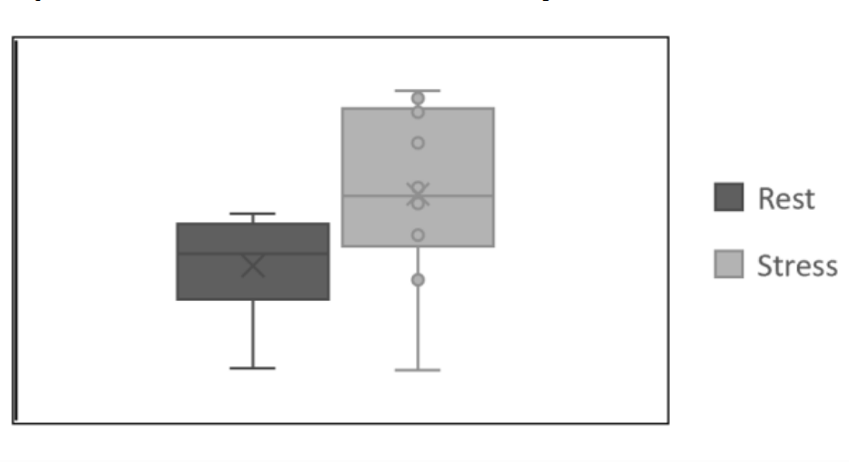
Abstract: Prolonged exposure to stress can have detrimental effects on physical and mental health. This study aimed to monitor anxiety levels with minimal sensors by using trapezius muscle EMG and ECG data during a stress task. Ten healthy adults were recruited, and HRV features were compared with EEG to determine alertness during the stress task. EMG data was compared with EEG β and α wave activity to determine stress levels. During the stress task, an increased β to α ratio in EEG indicated heightened alertness and focus, with a significant increase in trapezius muscle activity and asymmetry to indicate increased muscle tension and stress. Trapezius muscle EMG did prove to be an effective method for determining stress when compared to EEG. ECG derived HRV features showed potential to determine stress levels but require further testing. Based on the results, trapezius muscle EMG and ECG can potentially quantify stress and relate anxiety levels from EEG.
Publication Thumbnail

Ahmed, M., Kaya, M., Taebi, A., Gamage, P.T. (2023).
The Role of Meditation in Stress Recovery and Performance: An EEG Study.
ASME International Mechanical Engineering Conference and Exposition, New Orleans, LA, V005T06A029.
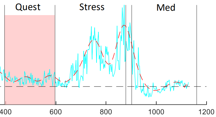
Abstract: This study aimed to investigate the effects of meditation on recovery time following a stressor and cognitive performance. Two sessions were performed in this study, one as a control without participants meditating prior to the stress task and one with meditation. EEG signals were recorded from 24 channels and processed using frequency domain analysis to measure power in various frequency bands. Participants for this pilot study were 10 healthy adults between the ages of 18 and 35. Reaction time and response accuracy were used as performance measures during the stressor with slower reaction times indicating lower levels of alertness. Brain activity prior to the stressor was compared with activity following the stress task, and the time needed to return to baseline was observed. The effect of meditation on recovery time and performance was analyzed and a gradual return to baseline values was observed. Recovery time varied among participants, with some taking longer than others to recover but no significant change was observed with meditation. Performance-wise, participants who meditated prior to the stressor had improved reaction time and accuracy when compared to the control group. The results suggest that interventions such as meditation or mindfulness practices may help individuals improve performance, but further research is needed to confirm these findings.
Publication Thumbnail

Rahman, M.M., Taebi, A. (2023).
Reconstruction of 3-Axis Seismocardiogram from Right-to-left and Head-to-foot Components Using A Long Short-Term Memory Network.
IEEE 19th International Conference on Body Sensor Networks, Boston, MA.
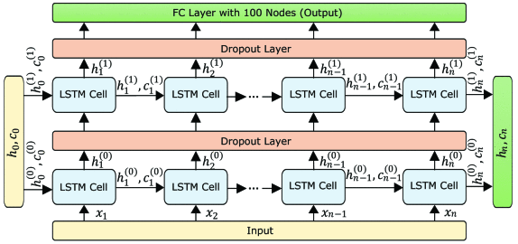
Abstract: This pilot study aims to develop a deep learning model for predicting seismocardiogram (SCG) signals in the dorsoventral direction from the SCG signals in the right-to-left and head-to-foot directions (SCGx and SCGy). The dataset used for the training and validation of the model was obtained from 15 healthy adult subjects. The SCG signals were recorded using tri-axial accelerometers placed on the chest of each subject. The signals were then segmented using electrocardiogram R waves, and the segments were downsampled, normalized, and centered around zero. The resulting dataset was used to train and validate a long short-term memory (LSTM) network with two layers and a dropout layer to prevent overfitting. The network took as input 100-time steps of SCGx and SCGy, representing one cardiac cycle, and outputted a vector that mapped to the target variable being predicted. The results showed that the LSTM model had a mean square error of 0.09 between the predicted and actual SCG segments in the dorsoventral direction. The study demonstrates the potential of deep learning models for reconstructing 3-axis SCG signals using the data obtained from dual-axis accelerometers.
Publication Thumbnail

Rahman, M.M., Cook, J., Taebi, A. (2023).
Non-contact heart vibration measurement using computer vision-based seismocardiography.
Scientific Reports, 13, 11787.
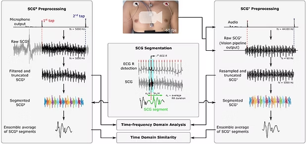
Abstract: Seismocardiography (SCG) is the noninvasive measurement of local vibrations of the chest wall produced by the mechanical activity of the heart and has shown promise in providing clinical information for certain cardiovascular diseases including heart failure and ischemia. Conventionally, SCG signals are recorded by placing an accelerometer on the chest. In this paper, we propose a novel contactless SCG measurement method to extract them from chest videos recorded by a smartphone. Our pipeline consists of computer vision methods including the Lucas–Kanade template tracking to track an artificial target attached to the chest, and then estimate the SCG signals from the tracked displacements. We evaluated our pipeline on 14 healthy subjects by comparing the vision-based SCGv estimations with the gold-standard SCGg measured simultaneously using accelerometers attached to the chest. The similarity between SCGg and SCGv was measured in the time and frequency domains using the Pearson correlation coefficient, a similarity index based on dynamic time warping (DTW), and wavelet coherence. The average DTW-based similarity index between the signals was 0.94 and 0.95 in the right-to-left and head-to-foot directions, respectively. Furthermore, SCGv signals were utilized to estimate the heart rate, and these results were compared to the gold-standard heart rate obtained from ECG signals. The findings indicated a good agreement between the estimated heart rate values and the gold-standard measurements (bias = 0.649 beats/min). In conclusion, this work shows promise in developing a low-cost and widely available method for remote monitoring of cardiovascular activity using smartphone videos.
Publication Thumbnail

Mann, A., Cook, J., Umar, M., Khalili, F., Taebi, A. (2022).
Heart Rate Monitoring Using Heart Acoustics.
ASME International Mechanical Engineering Conference and Exposition, Columbus, OH, V004T05A069.
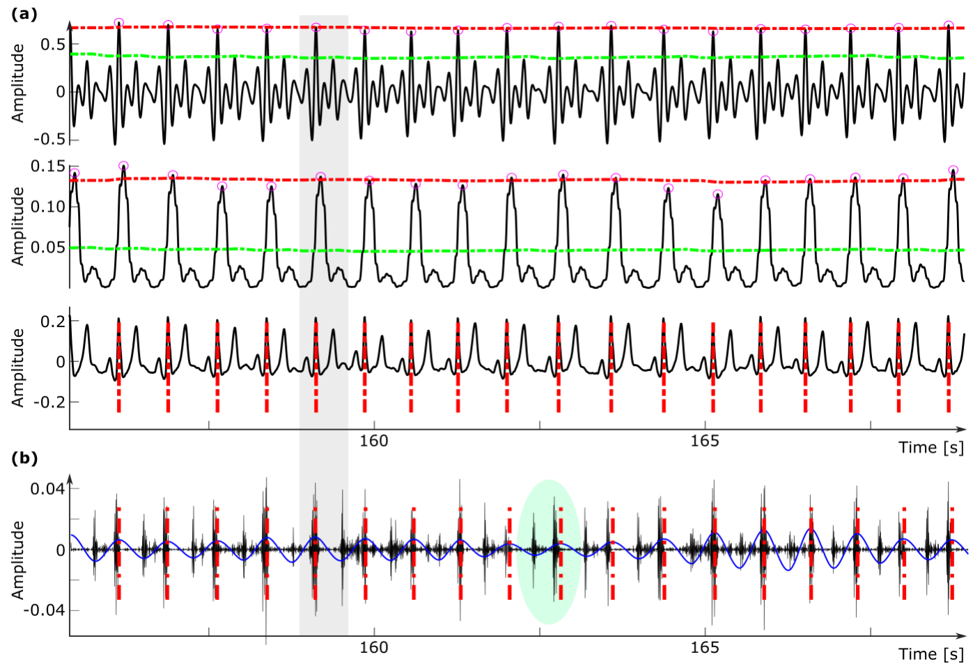
Abstract: Cardiovascular diseases (CVDs) are the leading cause of death in the United States. In many cases, CVDs go unnoticed or are diagnosed late, contributing to the high death rate of such diseases. To address this issue, new methods for the early diagnosis of CVDs should be developed. In many medical conditions, heart rate can play an important role as an early indicator of heart diseases. In this pilot study, a heart rate monitoring method based on cardiovascular-induced sounds is investigated. For this purpose, phonocardiography (PCG) signals are measured noninvasively on the body surface of five healthy subjects (21–24 years) using an electronic stethoscope. In addition, electrocardiography (ECG) was used as a gold-standard method of cardiac monitoring. The PCG signals were then post-processed using custom-built algorithms to estimate the subject heart rate. These estimated heart rates were then compared with the heart rate calculated from the ECG signal using the well-known Pan-Tompkins algorithm. Results showed that the heart rate estimations from the acoustic modalities were consistent with those calculated from the gold-standard ECG.
Publication Thumbnail

Taebi, A. (2022).
Deep Learning for Computational Hemodynamics: A Brief Review of Recent Advances.
Fluids, 7(6): 197.
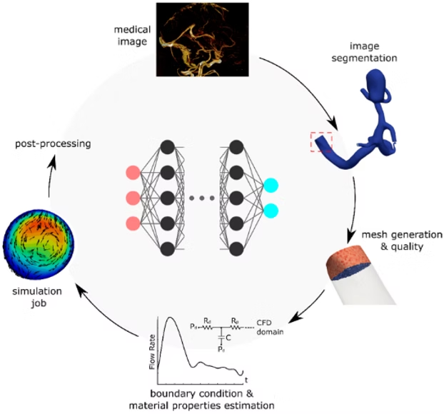
Abstract: Computational fluid dynamics (CFD) modeling of blood flow plays an important role in better understanding various medical conditions, designing more effective drug delivery systems, and developing novel diagnostic methods and treatments. However, despite significant advances in computational technology and resources, the expensive computational cost of these simulations still hinders their transformation from a research interest to a clinical tool. This bottleneck is even more severe for image-based, patient-specific CFD simulations with realistic boundary conditions and complex computational domains, which make such simulations excessively expensive. To address this issue, deep learning approaches have been recently explored to accelerate computational hemodynamics simulations. In this study, we review recent efforts to integrate deep learning with CFD and discuss the applications of this approach in solving hemodynamics problems, such as blood flow behavior in aorta and cerebral arteries. We also discuss potential future directions in the field. In this review, we suggest that incorporating physiologic understandings and underlying fluid mechanics laws in deep learning models will soon lead to a paradigm shift in the development novel non-invasive computational medical decisions.
Editor's Choice
Publication Thumbnail

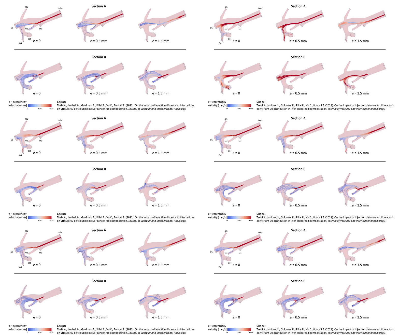
Taebi, A., Janibek, N., Goldman, R., Pillai, R., Vu, C., Roncali, E. (2022).
The impact of injection distance to bifurcations on yttrium-90 distribution in liver cancer radioembolization.
Journal of Vascular and Interventional Radiology, 33(6): 668-677e1.
Abstract: Purpose: To model the effect of the injection location on the distribution of yttrium-90 (90Y) microspheres in the liver during radioembolization using computational simulation and to determine the potential effects of radial movements of the catheter tip. Materials and Methods: Numerical studies were conducted using images from a representative patient with hepatocellular carcinoma. The right hepatic artery (RHA) was segmented from contrast-enhanced cone-beam computed tomography scans. The blood flow was investigated in the trunk of the RHA using numerical simulations for 6 injection position scenarios at 2 sites located at a distance of approximately 5 and 20 mm upstream of the first bifurcation (RHA diameters of approximately 4.6 mm). The 90Y delivery to downstream vessels was calculated from the simulated hepatic artery hemodynamics. Results: Varying the injection location along the RHA and across the vessel cross-section resulted in different simulated microsphere distributions in the downstream vascular bed. When the catheter tip was 5 mm upstream of the bifurcation, 90Y distribution in the downstream branches varied by as much as 53% with a 1.5-mm radial movement of the tip. However, the catheter radial movement had a weaker effect on the microsphere distribution when the injection plane was farther from the first bifurcation (20 mm), with a maximum delivery variation of 9% to a downstream branch. Conclusions: An injection location far from bifurcations is recommended to minimize the effect of radial movements of the catheter tip on the microsphere distribution.
featured CME JVIR article of the month for June 2022
Publication Thumbnail

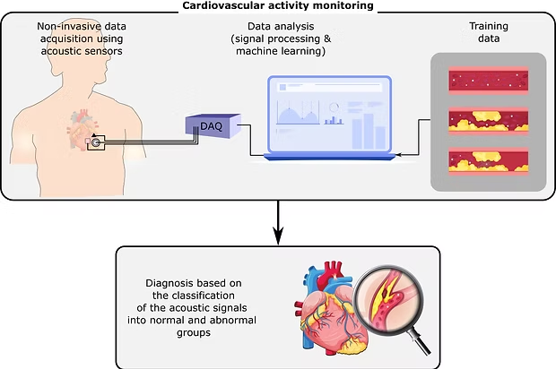
Cook, J., Umar, M., Khalili, F., Taebi, A. (2022).
Body acoustics for the non-invasive diagnosis of medical conditions.
Bioengineering, 9(4): 149.
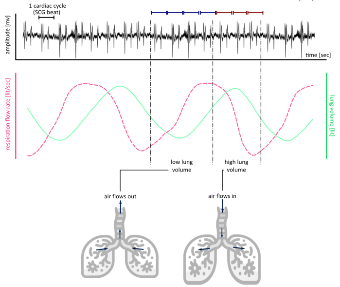
Abstract: In the past few decades, many non-invasive monitoring methods have been developed based on body acoustics to investigate a wide range of medical conditions, including cardiovascular diseases, respiratory problems, nervous system disorders, and gastrointestinal tract diseases. Recent advances in sensing technologies and computational resources have given a further boost to the interest in the development of acoustic-based diagnostic solutions. In these methods, the acoustic signals are usually recorded by acoustic sensors, such as microphones and accelerometers, and are analyzed using various signal processing, machine learning, and computational methods. This paper reviews the advances in these areas to shed light on the state-of-the-art, evaluate the major challenges, and discuss future directions. This review suggests that rigorous data analysis and physiological understandings can eventually convert these acoustic-based research investigations into novel health monitoring and point-of-care solutions.
Publication Thumbnail

Taebi, A., Khalili, F. (2021).
Advances in Noninvasive Diagnosis Based on Body Sounds and Vibrations.
ASME International Mechanical Engineering Conference and Exposition, V005T05A080.

Abstract: This paper provides a brief overview of the advances in the area of early identification of different types of abnormalities and diseases, including respiratory illnesses and cardiovascular diseases, using noninvasive screening of biomedical acoustic signals. These signals include sounds and vibrations generated by different human body organs and systems that can be measured on the body surface using sensors such as stethoscopes and accelerometers. In this study, the measurement methods and signal processing algorithms for customized feature extraction and classification as well as clinical potentials, current limitations, and future directions are briefly reviewed and discussed.
Publication Thumbnail

Khalili, F., Taebi, A. (2021).
Advances in Computational Fluid Dynamics Modeling of the Body Sounds as a Noninvasive Diagnosis Method.
ASME International Mechanical Engineering Conference and Exposition, V005T05A041.
Abstract: This paper provides a concise overview of the recent advances in the computational fluid dynamics modeling of flow-induced sounds, a valuable non-invasive tool that delivers complementary information for the early detection of cardiovascular and pulmonary diseases. An abnormal flow through an unhealthy artery consists of turbulent pressure fluctuations that interact with the arterial walls, leading to the sound waves propagated through the surrounding tissue. These sound waves recorded on the epidermal surface are vascular sounds known as murmurs. Detailed studies of the adverse flow conditions associated with cardiovascular and pulmonary diseases are vital to enhance our understanding of the mechano-acoustics mechanisms of flow-induced sound sources. This information can lead us to predictive, non-invasive techniques for diagnosing different diseases such as atherosclerosis and aneurysm before they progress to severe cases. This necessity suggests that more studies are necessary to develop strategies that can be employed to detect cardiovascular diseases without the need for invasive approaches.
Publication Thumbnail

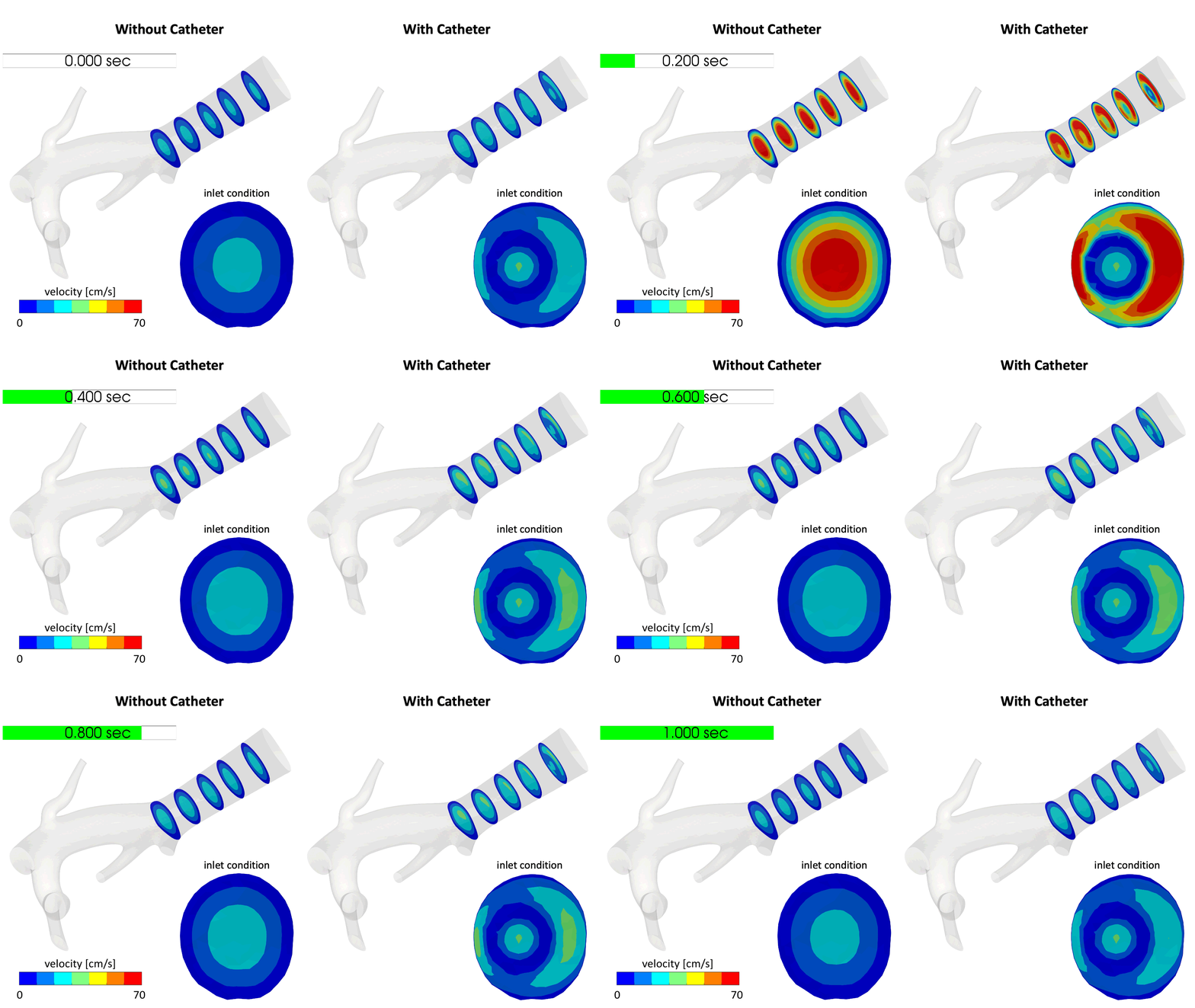
Taebi, A., Berk, S., Roncali, E. (2021).
Realistic boundary conditions in SimVascular through inlet catheter modeling.
BMC Research Notes, 14, 215.
Abstract: Objective: This study aims at developing a pipeline that provides the capability to include the catheter effect in the computational fluid dynamics (CFD) simulations of the cardiovascular system and other human vascular flows carried out with the open-source software SimVascular. This tool is particularly useful for CFD simulation of interventional radiology procedures such as tumor embolization where estimation of a therapeutic agent distribution is of interest. Results: A pipeline is developed that generates boundary condition files which can be used in SimVascular CFD simulations. The boundary condition files are modified such that they simulate the effect of catheter presence on the flow field downstream of the inlet. Using this pipeline, the catheter flow, velocity profile, radius, wall thickness, and deviation from the vessel center can be defined. Since our method relies on the manipulation of the boundary condition that is imposed on the inlet, it is sensitive to the mesh density. The finer the mesh is (especially around the catheter wall), the more accurate the velocity estimations are. In this study, we also utilized this pipeline to qualitatively investigate the effect of catheter presence on the flow field in a truncated right hepatic arterial tree of a liver cancer patient.
Publication Thumbnail

Taebi, A., Costa, G.C.A., Roncali, E. (2021).
Personalized dosimetry for brain cancer radioembolization: A feasibility study.
Journal of Nuclear Medicine 62 (supplement 1): 1582.

Publication Thumbnail

Khalili, F., Gamage, P.T., Taebi, A., Johnson, M.E., Roberts, R.B., Mitchell, J. (2021).
Spectral decomposition of the flow and characterization of the sound signals generated through stenoses of different levels of severity.
Bioengineering, 8(3): 41.
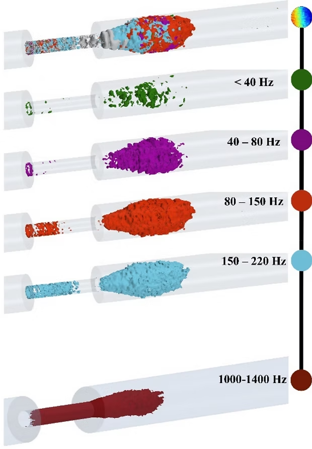
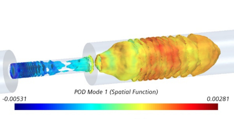
Abstract: Treatments of atherosclerosis depend on the severity of the disease at the diagnosis time. Non-invasive diagnosis techniques, capable of detecting stenosis at early stages, are essential to reduce associated costs and mortality rates. We used computational fluid dynamics and acoustics analysis to extensively investigate the sound sources arising from high-turbulent fluctuating flow through stenosis. The frequency spectral analysis and proper orthogonal decomposition unveiled the frequency contents of the fluctuations for different severities and decomposed the flow into several frequency bandwidths. Results showed that high-intensity turbulent pressure fluctuations appeared inside the stenosis for severities above 70%, concentrated at plaque surface, and immediately in the post-stenotic region. Analysis of these fluctuations with the progression of the stenosis indicated that (a) there was a distinct break frequency for each severity level, ranging from 40 to 230 Hz, (b) acoustic spatial-frequency maps demonstrated the variation of the frequency content with respect to the distance from the stenosis, and (c) high-energy, high-frequency fluctuations existed inside the stenosis only for severe cases. This information can be essential for predicting the severity level of progressive stenosis, comprehending the nature of the sound sources, and determining the location of the stenosis with respect to the point of measurements.
Publication Thumbnail

Khalili, F., Gamage, P.T., Taebi, A., Johnson, M.E., Roberts, R.B., Mitchell, J. (2021).
Spectral decomposition and sound source localization of highly disturbed flow through a severe arterial stenosis.
Bioengineering, 8(3): 34.
Abstract: For the early detection of atherosclerosis, it is imperative to explore the capabilities of new, effective noninvasive diagnosis techniques to significantly reduce the associated treatment costs and mortality rates. In this study, a multifaceted comprehensive approach involving advanced computational fluid dynamics combined with signal processing techniques was exploited to investigate the highly turbulent fluctuating flow through arterial stenosis. The focus was on localizing high-energy mechano-acoustic source potential to transmit to the epidermal surface. The flow analysis results showed the existence of turbulent pressure fluctuations inside the stenosis and in the post-stenotic region. After analyzing the turbulent kinetic energy and pressure fluctuations on the flow centerline and the vessel wall, the point of maximum excitation in the flow was observed around two diameters downstream of the stenosis within the fluctuating zone. It was also found that the concentration of pressure fluctuation closer to the wall was higher inside the stenosis compared to the post-stenotic region. Additionally, the visualization of the most energetic proper orthogonal decomposition (POD) mode and spectral decomposition of the flow indicated that the break frequencies ranged from 80 to 220 Hz and were correlated to the eddies generated within these regions.
Publication Thumbnail

Taebi, A., Vu, C.T., Roncali, E. (2021).
Multiscale computational fluid dynamics modeling for personalized liver cancer radioembolization dosimetry.
ASME Journal of Biomechanical Engineering, 143(1): 011002.
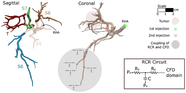
Abstract: Yttrium-90 (90Y) radioembolization is a minimally invasive procedure increasingly used for advanced liver cancer treatment. In this method, radioactive microspheres are injected into the hepatic arterial bloodstream to target, irradiate, and kill cancer cells. Accurate and precise treatment planning can lead to more efficient and safer treatment by delivering a higher radiation dose to the tumor while minimizing the exposure of the surrounding liver parenchyma. Treatment planning primarily relies on the estimated radiation dose delivered to tissue. However, current methods used to estimate the dose are based on simplified assumptions that make the dosimetry results unreliable. In this work, we present a computational model to predict the radiation dose from the 90Y activity in different liver segments to provide a more realistic and personalized dosimetry. Computational fluid dynamics (CFD) simulations were performed in a 3D hepatic arterial tree model segmented from cone-beam CT angiographic data obtained from a patient with hepatocellular carcinoma (HCC). The microsphere trajectories were predicted from the velocity field. 90Y dose distribution was then calculated from the volumetric distribution of the microspheres. Two injection locations were considered for the microsphere administration, a lobar and a selective injection. Results showed that 22% and 82% of the microspheres were delivered to the tumor, after each injection, respectively, and the combination of both injections ultimately delivered 49% of the total administered 90Y microspheres to the tumor. Results also illustrated the nonhomogeneous distribution of microspheres between liver segments, indicating the importance of developing patient-specific dosimetry methods for effective radioembolization treatment.
honorable mention for the 2022 Skalak Award, among top 3 papers published by JBME in 2021 & 2022: 10.1115/1.4065051
Publication Thumbnail

Taebi, A., Vu, C.T., Roncali, E. (2020).
Prediction of Blood Flow Distribution in Liver Radioembolization Using Convolutional Neural Networks.
ASME International Mechanical Engineering Conference and Exposition, Portland, OR, V005T05A036.
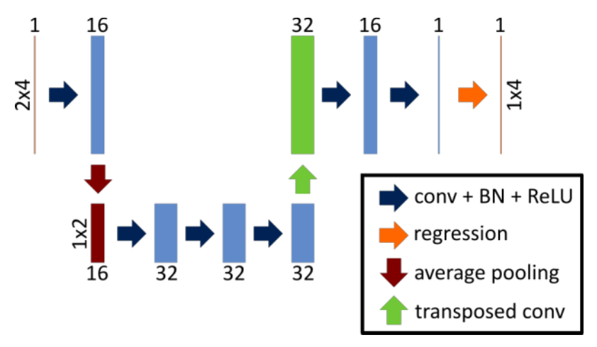
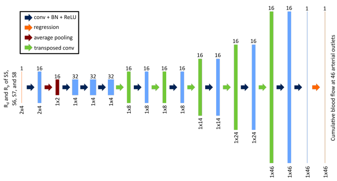
Abstract: We have developed a new dosimetry approach, called CFDose, for liver cancer radioembolization based on computational fluid dynamics (CFD) simulation in the hepatic arterial tree. Although CFDose overcomes some of the limitations of the current dosimetry methods such as the unrealistic assumption of homogeneous distribution of yttrium-90 in the liver, it suffers from the expensive computational cost of CFD simulations. To accelerate CFDose, we introduce a deep learning model to predict the blood flow distribution between the liver segments in a patient with hepatocellular carcinoma. The model was trained with the results of CFD simulations under different outlet boundary conditions. The model consisted of convolutional, average pooling and transposed convolution layers. A regression layer with a mean-squared-error loss function was utilized at the network output to estimate the arterial outlet blood flow. The mean-squared error and prediction accuracy were calculated to measure model performance. Results showed that the average difference between the CFD results and predicted flow data was less than 2.45% for all the samples in the test dataset. The proposed model thus estimated the blood flow distribution with high accuracy significantly faster than a CFD simulation. The network output can be used to estimate the yttrium-90 dose distribution in the liver in future studies.
Publication Thumbnail

Taebi, A., Vu, C.T., Roncali, E. (2020).
Estimation of Yttrium-90 Distribution in Liver Radioembolization using Computational Fluid Dynamics and Deep Neural Networks.
IEEE Engineering in Medicine and Biology Society, Montreal, QC, Canada, pp. 4974-4977.

Abstract: Yttrium-90 (90Y) radioembolization is a liver cancer therapy based on 90Y microspheres injected into the hepatic artery. Current dosimetry methods used to estimate the absorbed dose in order to prescribe the 90Y activity to inject are not accurate, which can affect the treatment effectiveness. A new dosimetry based on the hemodynamics simulation of the hepatic arterial tree, CFDose, aimed at overcoming some of the limitations of the current methods. However, due to the expensive computational cost of computational fluid dynamics (CFD) simulations, this method needs to be accelerated before it can be used in real-time during treatment planning. In this paper, we introduce a convolutional neural network model trained with the CFD results of a patient with hepatocellular carcinoma to predict the 90Y distribution under different downstream vasculature resistance conditions. The model performance was evaluated using two metrics, the mean squared error and prediction accuracy. The prediction accuracy showed that the average difference between the actual and predicted data was less than 1%. The proposed model could estimate the 90Y distribution significantly faster than a CFD simulation.
Publication Thumbnail

Taebi, A., Pillai, R., Roudsari, B., Vu, C., Roncali, E. (2020).
Computational modeling of the liver arterial blood flow for microsphere therapy: Effect of boundary conditions.
Bioengineering, 7(3): 64.
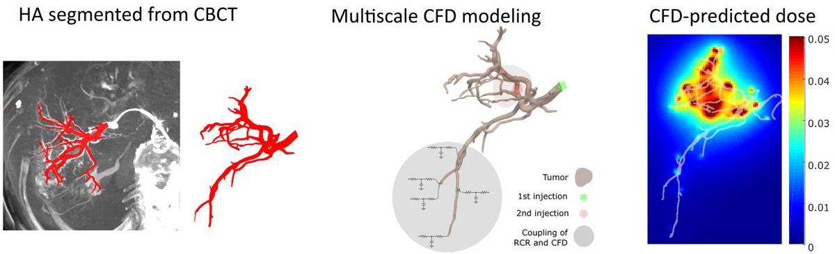
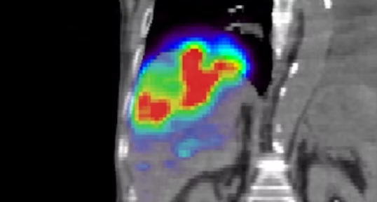
Abstract: Transarterial embolization is a minimally invasive treatment for advanced liver cancer using microspheres loaded with a chemotherapeutic drug or radioactive yttrium-90 (90Y) that are injected into the hepatic arterial tree through a catheter. For personalized treatment, the microsphere distribution in the liver should be optimized through the injection volume and location. Computational fluid dynamics (CFD) simulations of the blood flow in the hepatic artery can help estimate this distribution if carefully parameterized. An important aspect is the choice of the boundary conditions imposed at the inlet and outlets of the computational domain. In this study, the effect of boundary conditions on the hepatic arterial tree hemodynamics was investigated. The outlet boundary conditions were modeled with three-element Windkessel circuits, representative of the downstream vasculature resistance. Results demonstrated that the downstream vasculature resistance affected the hepatic artery hemodynamics such as the velocity field, the pressure field and the blood flow streamline trajectories. Moreover, the number of microspheres received by the tumor significantly changed (more than 10% of the total injected microspheres) with downstream resistance variations. These findings suggest that patient-specific boundary conditions should be used in order to achieve a more accurate drug distribution estimation with CFD in transarterial embolization treatment planning.
featured on the journal's website
Publication Thumbnail

Roncali, E., Taebi, A., Spencer, B., Costa, G.C.A., Rusnak, M., Caudle, D., Roudsari, B., Pillai, R., Foster, C., Vu, C. (2020).
Comparison of Y-90 liver dose distribution predicted with fluid dynamics with Y-90 PET.
Journal of Nuclear Medicine 61 (supplement 1): 1308.

Publication Thumbnail

Roncali, E.,Taebi, A., Roudsari, B.S., Vu, C.T. (2020).
Personalized dosimetry for liver cancer radioembolization based on computational fluid dynamics.
Annals of Biomedical Engineering, 48(5): 1499-1510.
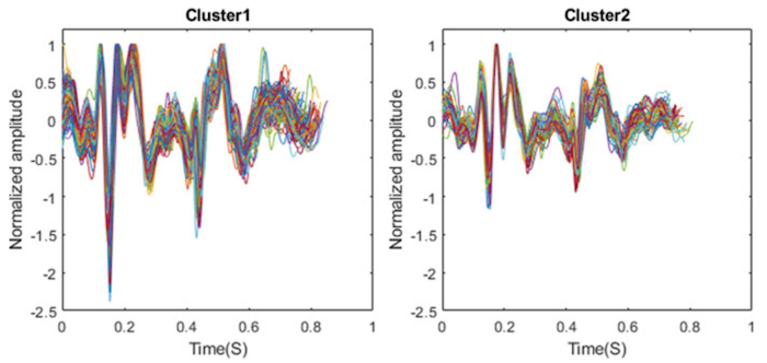
Abstract: Yttrium-90 (Y-90) transarterial radioembolization uses radioactive microspheres injected into the hepatic artery to irradiate liver tumors internally. One of the major challenges is the lack of reliable dosimetry methods for dose prediction and dose verification. We present a patient-specific dosimetry approach for personalized treatment planning based on computational fluid dynamics (CFD) simulations of the microsphere transport combined with Y-90 physics modeling called CFDose. The ultimate goal is the development of a software to optimize the amount of activity and injection point for optimal tumor targeting. We present the proof-of-concept of a CFD dosimetry tool based on a patient’s angiogram performed in standard-of-care planning. The hepatic arterial tree of the patient was segmented from the cone-beam CT (CBCT) to predict the microsphere transport using multiscale CFD modeling. To calculate the dose distribution, the predicted microsphere distribution was convolved with a Y-90 dose point kernel. Vessels as small as 0.45 mm were segmented, the microsphere distribution between the liver segments using flow analysis was predicted, the volumetric microsphere and resulting dose distribution in the liver volume were computed. The patient was imaged with positron emission tomography (PET) 2 h after radioembolization to evaluate the Y-90 distribution. The dose distribution was found to be consistent with the Y-90 PET images. These results demonstrate the feasibility of developing a complete framework for personalized Y-90 microsphere simulation and dosimetry using patient-specific input parameters.
Publication Thumbnail

Gamage, P.T., Azad, M.K., Taebi, A., Sandler, R.H., Mansy, H.A. (2020).
Clustering of SCG Events using Unsupervised Machine Learning.
In book: Signal Processing in Medicine and Biology. Springer, Cham.
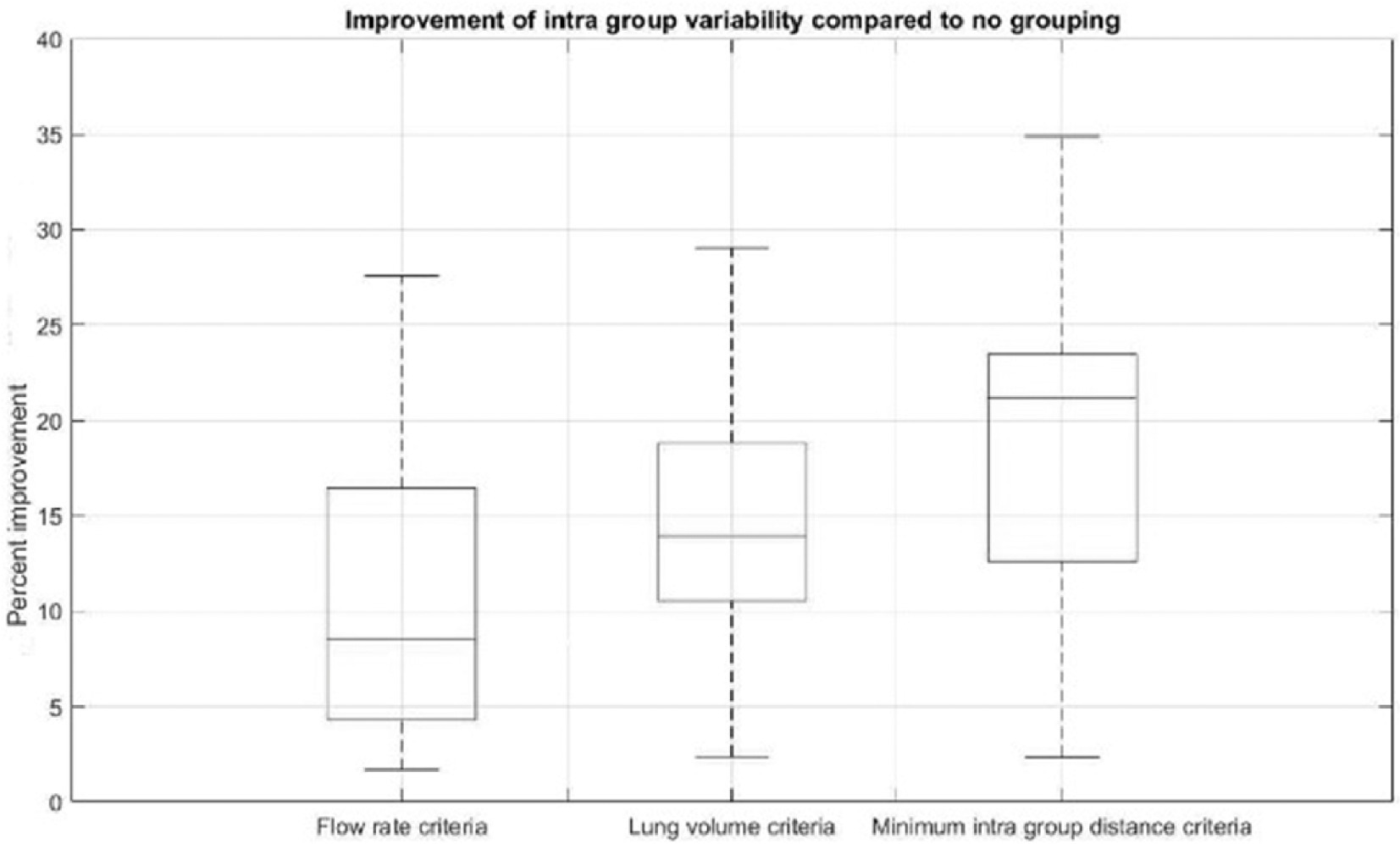
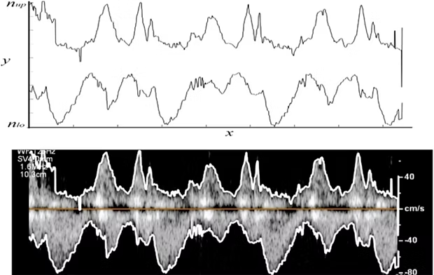
Abstract: Seismocardiography (SCG) is the measurement of chest surface vibrations induced by cardiac activity. SCG beats are typically averaged to reduce noise and determine average SCG waveforms and features. Variability in SCG morphology impedes precise determination of average waveforms. Hence, it is desirable to group SCG beats into clusters with minimal intra-cluster heterogeneity. Cardio-pulmonary interactions are known to contribute to SCG variability. Therefore, grouping SCG signals by their respiratory phase may be helpful. SCG signals and respiratory flowrate were simultaneously measured in seventeen subjects (Age: 23 ± 3.5 years, 7 female). Unsupervised machine learning was implemented to cluster SCG beats according to their morphology. The time domain amplitudes of the SCG beats were used as the feature vector. K-medoids clustering was employed with dynamic time warping (DTW) distance as the heterogeneity measure. The quality of the clustering was measured using mean silhouette values and the elbow method for varying clusters numbers. Optimal clustering was achieved when SCG beats were split into two groups. Using respiratory flow information, SCG beats were labeled as inspiratory vs. expiratory, and as high vs. low lung volumes. The SCG groups determined by machine learning were compared with these labels. Grouping SCG based on lung volume phases yielded more homogeneous clusters than grouping by inspiration vs. expiration (p < 0.01). Unsupervised clustering reduced the intra-cluster variability by an average of 15% across subjects. Grouping by lung volume and inspiration vs. expiration reduced variability by 6% and 3%, respectively. The variability reduction may help more precise determination of average SCG waveforms and features, thereby improving SCG diagnostic utility.
Publication Thumbnail

Roncali, E., Taebi, A., Rusnak, M., Spencer, B., Caudle, D., Foster, C., Vu, C.T. (2019).
Personalized dosimetry for liver cancer radioembolization using computational fluid dynamics.
European Journal of Nuclear Medicine and Molecular Imaging 46 (Suppl 1): S134.

Publication Thumbnail

Sandler, R.H., Azad, M.K., Rahman, B., Taebi, A., Gamage, P., Raval, N., Mentz, R.J., Mansy, H.A. (2019).
Minimizing Seismocardiography Variability by Accounting for Respiratory Effects.
Journal of Cardiac Failure 25(8): S172.

Publication Thumbnail

Taebi, A., Sandler, R.H., Kakavand, B., Mansy, H.A. (2019).
Extraction of Peak Velocity Profiles from Doppler Echocardiography Using Image Processing.
Bioengineering, 6(3): 64.
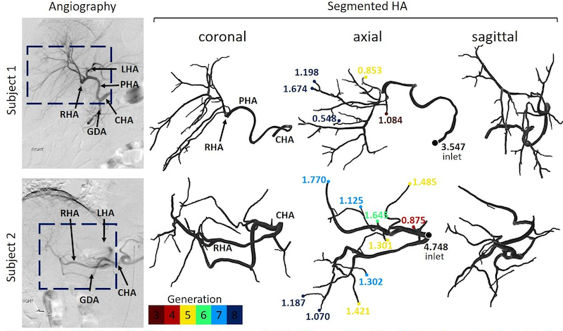
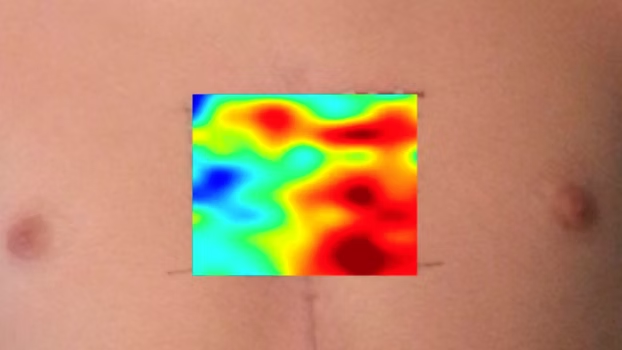
Abstract: The objective of this study is to extract positive and negative peak velocity profiles from Doppler echocardiographic images. These profiles are currently estimated using tedious manual approaches. Profiles can be used to establish realistic boundary conditions for computational hemodynamic studies and to estimate cardiac time intervals, which are of clinical utility. In the current study, digital image processing algorithms that rely on intensity calculations and two different thresholding methods were proposed and tested. Image intensity histograms were used to guide threshold choices, which were selected such that the resulting velocity profiles appropriately represent Doppler shift envelopes. The resulting peak velocity profiles contained artifacts in the form of sudden velocity changes and possible outliers. To reduce these artifacts, image smoothing using a moving average process was then implemented. Bland–Altman analysis suggested good agreement between the two thresholding methods. Artifacts decreased when image smoothing was performed. Results also suggested that one thresholding method tended to provide the lower limit (i.e., underestimate) of velocities, while the second tended to provide the velocity upper limit (i.e., overestimate). Combining estimates from both methods appeared to provide a smoother peak velocity profile estimate. The proposed automated approach may be useful for objective estimation of peak velocity profiles, which may be helpful for computational hemodynamic studies and may increase the efficiency of current clinical diagnostic tools.
Publication Thumbnail

Taebi, A., Roudsari, B.S., Vu, C., Cherry, S.R., Roncali, E. (2019).
Hepatic arterial tree segmentation: Towards patient-specific dosimetry for liver cancer radioembolization.
Journal of Nuclear Medicine 60 (supplement 1) 122.

Publication Thumbnail

Taebi, A., Solar, B.E., Bomar, A.J., Sandler, R.H., Mansy, H.A. (2019).
Recent Advances in Seismocardiography.
Vibration, 2(1), 64-86.
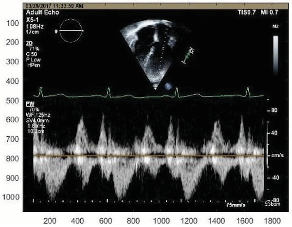
Abstract: Cardiovascular disease is a major cause of death worldwide. New diagnostic tools are needed to provide early detection and intervention to reduce mortality and increase both the duration and quality of life for patients with heart disease. Seismocardiography (SCG) is a technique for noninvasive evaluation of cardiac activity. However, the complexity of SCG signals introduced challenges in SCG studies. Renewed interest in investigating the utility of SCG accelerated in recent years and benefited from new advances in low-cost lightweight sensors, and signal processing and machine learning methods. Recent studies demonstrated the potential clinical utility of SCG signals for the detection and monitoring of certain cardiovascular conditions. While some studies focused on investigating the genesis of SCG signals and their clinical applications, others focused on developing proper signal processing algorithms for noise reduction, and SCG signal feature extraction and classification. This paper reviews the recent advances in the field of SCG.
all time most cited paper of the journal
Publication Thumbnail

Taebi, A., Sandler R.H., Kakavand, B., Mansy, H.A. (2018).
Estimating Peak Velocity Profiles from Doppler Echocardiography using Digital Image Processing.
IEEE Signal Processing in Medicine and Biology, Philadelphia, PA.
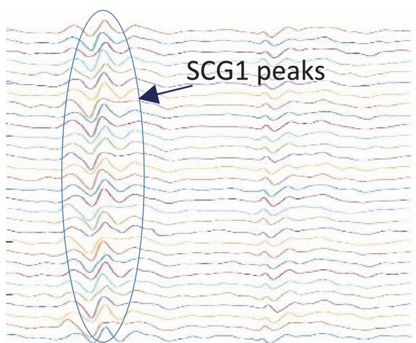
Abstract: This study aims at developing a digital signal processing algorithm to extract positive and negative peak velocity profiles from Doppler echocardiographic images. These profiles are useful in estimating cardiac time intervals and establishing realistic boundary conditions for computational hemodynamic studies. The proposed image processing algorithm is based on two different thresholding methods. The histograms of image intensity function were used to help threshold values selection so that the algorithm yields velocity profiles properly represent Doppler shift envelopes. One of the thresholding methods tended to provide the lower-limit (i.e. underestimate) of the velocity profile, while the second tended to provide the upper-limit of the velocity profile (i.e., overestimate). The final peak velocity profiles were estimated from the combination of the estimates from both thresholding methods. The peak velocity profiles were then qualitatively compared with the results of the standard edge detection methods such as Canny and Prewitt approximations. The proposed automated approach might be helpful for objective estimation of peak velocities and cardiac time intervals.
Publication Thumbnail

Gamage, P.T., Azad, M.K., Taebi, A., Sandler, R.H., Mansy, H.A. (2018).
Clustering Seismocardiographic Events using Unsupervised Machine Learning.
IEEE Signal Processing in Medicine and Biology, Philadelphia, PA.
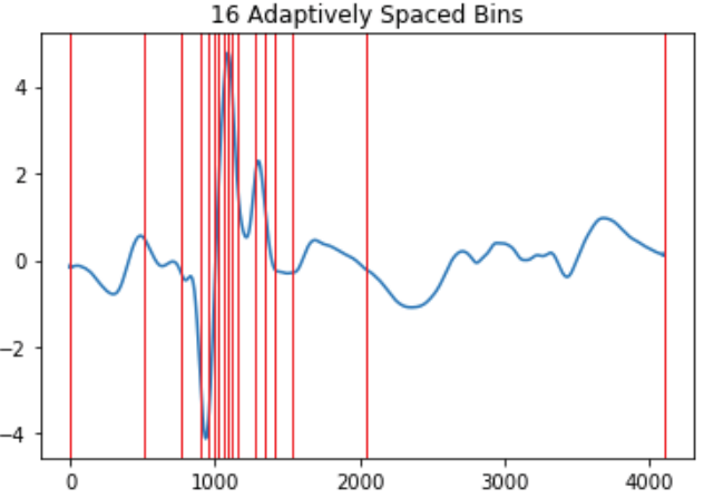
Abstract: Seismocardiographic (SCG) signal morphology is known to be affected by cardio-pulmonary interactions, which introduce variability in the SCG signal. Hence, grouping of SCG signals according to their respiratory phase can reduce their morphological dissimilarity. In addition, correlating SCG with pulmonary phases may provide more insights into the nature of cardio-pulmonary interactions. This study uses unsupervised machine learning to cluster SCG events based on their morphology. Here, K-means clustering was employed using the time domain amplitude as the feature vector. The method is applied on measured SCG data from 5 male subjects (Age: 30 ± 5.8 years). The mean Silhouette values for different number of clusters suggested that optimal clustering was reached when SCG waveforms were divided into two groups. Using respiratory flow information, SCG waves were labeled as inspiratory vs. expiratory or high vs. low lung volume. The SCG clusters were then compared with these labels and purity values were calculated. The distributions of clustered SCG events in relation to respiratory flowrate and lung volume phases showed consistent trends in all subjects. Results suggested that grouping SCG based on lung volume phases would yield more homogeneous groups and, hence, would keep SCG variability (within each group) to a minimum. The demonstrated utility of the proposed machine learning approach in identifying respiratory phases from SCG waveforms may obviate the need for simultaneous respiratory measurements.
Publication Thumbnail

Taebi, A., Solar, B.E., Mansy, H.A. (2018).
An Adaptive Feature Extraction Algorithm for Classification of Seismocardiographic Signals.
IEEE SoutheastCon, Saint Petersburg, FL.
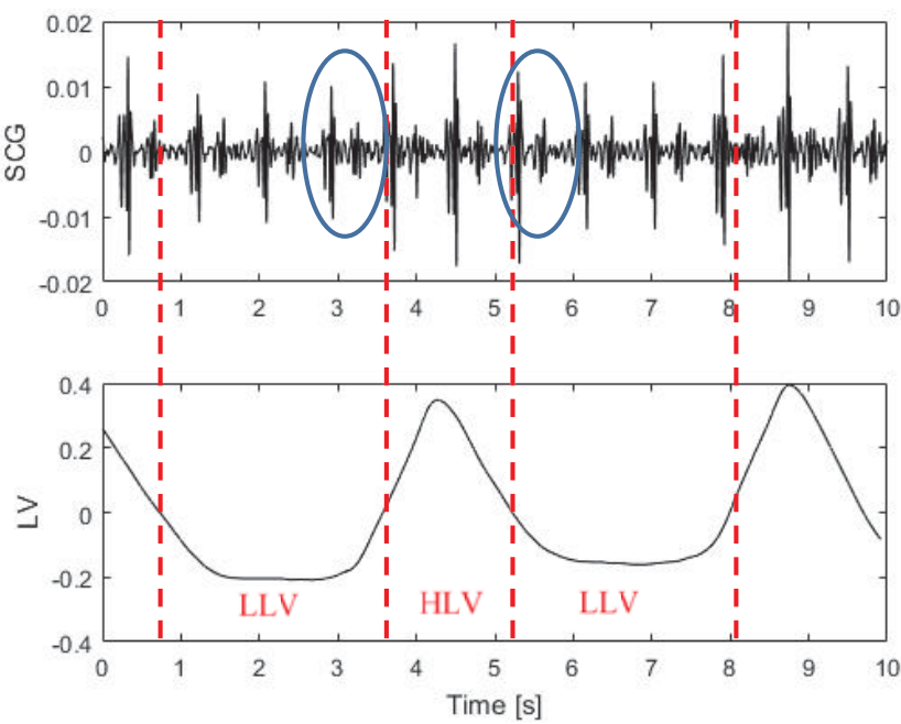
Abstract: This paper proposes a novel adaptive feature extraction algorithm for seismocardiographic (SCG) signals. The proposed algorithm divides the SCG signal into a number of bins., where the length of each bin is determined based on the signal change within that bin. For example., when the signal variation is steeper., the bins are shorter and vice versa. The proposed algorithm was used to extract features of the SCG signals recorded from 7 healthy individuals (Age: 29.4±4.5 years) during different lung volume phases. The output of the feature extraction algorithm was fed into a support vector machines classifier to classify SCG events into two classes of high and low lung volume (HLV and LLV). The classification results were compared with currently available non-adaptive feature extraction methods for different number of bins. Results showed that the proposed algorithm led to a classification accuracy of ~90%. The proposed algorithm outperformed the non-adaptive algorithm., especially as the number of bins was reduced. For example, for 16 bins, F1 score for the adaptive and non-adaptive methods were 0.91±0.05 and 0.63±0.08., respectively.
Publication Thumbnail

Taebi, A., Bomar, A.J., Sandler, R.H., Mansy, H.A. (2018).
Heart Rate Monitoring During Different Lung Volume Phases Using Seismocardiography.
IEEE SoutheastCon, Saint Petersburg, FL.
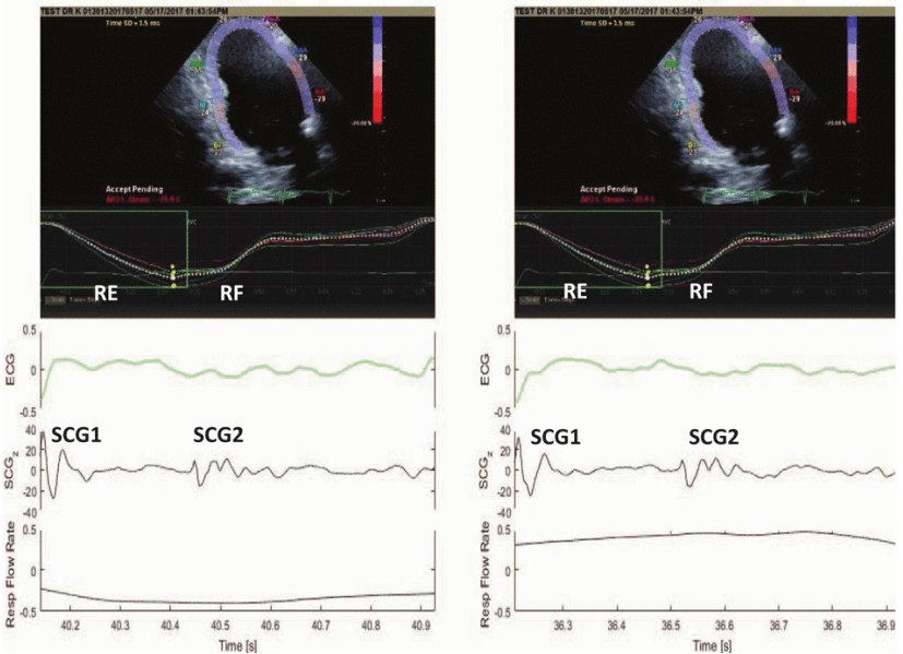
Abstract: Seismocardiography (SCG) is a non-invasive method that can be used for cardiac activity monitoring. This paper presents a new electrocardiogram (ECG) independent approach for estimating heart rate (HR) during low and high lung volume (LLV and HLV, respectively) phases using SCG signals. In this study, SCG, ECG, and respiratory flow rate (RFR) signals were measured simultaneously in 7 healthy subjects. The lung volume information was calculated from the RFR and was used to group the SCG events into low and high lung-volume groups. LLV and HLV SCG events were then used to estimate the subjects HR as well as the HR during LLV and HLV in 3 different postural positions, namely supine, 45 degree heads-up, and sitting. The performance of the proposed algorithm was tested against the standard ECG measurements. Results showed that the HR estimations from the SCG and ECG signals were in a good agreement (bias of 0.08 bpm). All subjects were found to have a higher HR during HLV (HRHLV) compared to LLV (HRLLV) at all postural positions. The HRHLV/HRLLV ratio was 1.11±0.071.08±0.05, 1.09±0.04, and 1.09±0.04 (mean±SD)) for supine, 45 degree-first trial, 45 degree-second trial, and sitting positions, respectively. This heart rate variability may be due, at least in part, to the well-known respiratory sinus arrhythmia. HR monitoring from SCG signals might be used in different clinical applications including wearable cardiac monitoring systems.
Publication Thumbnail

Taebi, A., Sandler, R.H., Kakavand, B., Mansy, H.A. (2017).
Seismocardiographic Signal Timing with Myocardial Strain.
IEEE Signal Processing in Medicine and Biology, Philadelphia, PA.

Abstract: Speckle Tracking Echocardiography (STE) is a relatively new method for cardiac function evaluation. In the current study, STE was used to investigate the timing of heart-induced mostly subaudible (i.e., below the frequency limit of human hearing) chest-wall vibrations in relation to the longitudinal myocardial strain. Such an approach may help elucidate the genesis of these vibrations, thereby improving their diagnostic value.
Publication Thumbnail

Solar, B.E., Taebi, A., Mansy, H.A. (2017).
Classification of Seismocardiographic Cycles into Lung Volume Phases.
IEEE Signal Processing in Medicine and Biology, Philadelphia, PA.
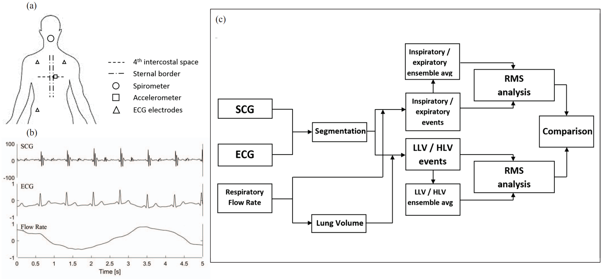
Abstract: In this study, a machine learning algorithm was developed to classify seismocardiographic (SCG) signals occurring during low and high lung volumes. The results demonstrated that morphological differences can be observed in SCG waveforms during respiration. SCG events were classified using a Radial Basis Function (RBF) support vector machine (SVM) algorithm into the two classes of low and high lung volume. Classification accuracy was found to be about 75%.
Publication Thumbnail

Taebi, A., Mansy, H.A. (2017).
Grouping Similar Seismocardiographic Signals Using Respiratory Information.
IEEE Signal Processing in Medicine and Biology, Philadelphia, PA.
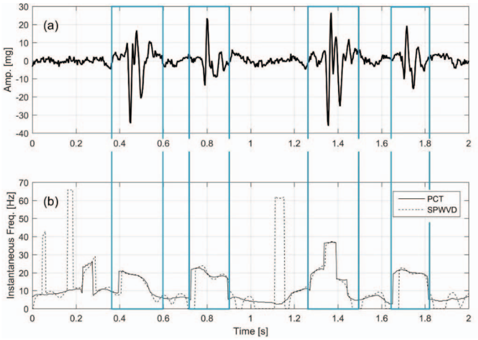
Abstract: Seismocardiography (SCG) offers a potential noninvasive method for cardiac monitoring. Quantification of the effects of different physiological conditions on SCG can lead to enhanced understanding of SCG genesis, and may explain how some cardiac pathologies may affect SCG morphology. In this study, the effect of the respiration on the SCG signal morphology is investigated. SCG, ECG, and respiratory flow rate signals were measured simultaneously in 7 healthy subjects. Results showed that SCG events tended to have two slightly different morphologies. The respiratory flow rate and lung volume information were used to group the SCG events into inspiratory/expiratory groups or low/high lung-volume groups, respectively. Although respiratory flow information could separate similar SCG events into two different groups, the lung volume information provided better grouping of similar SCGs. This suggests that variations in SCG morphology may be due, at least in part, to changes in the intrathoracic pressure or heart location since those parameters correlates more with lung volume than respiratory flow. Categorizing SCG events into different groups containing similar events allows more accurate estimation of SCG features, and better signal characterization, and classification.
Publication Thumbnail

Taebi, A., Mansy, H.A. (2017).
Analysis of Seismocardiographic Signals Using Polynomial Chirplet Transform and Smoothed Pseudo Wigner-Ville Distribution.
IEEE Signal Processing in Medicine and Biology, Philadelphia, PA.
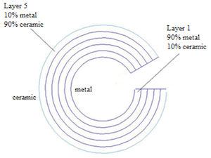
Abstract: Seismocardiographic (SCG) signals are chest surface vibrations induced by cardiac activity. These signals may offer a method for diagnosing and monitoring heart function. Successful classification of SCG signals in health and disease depends on accurate signal characterization and feature extraction. One approach of determining signal features is to estimate its time-frequency characteristics. In this regard, four different time-frequency distribution (TFD) approaches were used including short-time Fourier transform (STFT), polynomial chirplet transform (PCT), Wigner-Ville distribution (WVD), and smoothed pseudo Wigner-Ville distribution (SPWVD). Synthetic SCG signals with known time-frequency properties were generated and used to evaluate the accuracy of the different TFDs in extracting SCG spectral characteristics. Using different TFDs, the instantaneous frequency (IF) of each synthetic signal was determined and the error (NRMSE) in estimating IF was calculated. STFT had lower NRMSE than WVD for synthetic signals considered. PCT and SPWVD were, however, more accurate IF estimators especially for the signal with time-varying frequencies. PCT and SPWVD also provided better discrimination between signal frequency components. Therefore, the results of this study suggest that PCT and SPWVD would be more reliable methods for estimating IF of SCG signals. Analysis of actual SCG signals showed that these signals had multiple spectral components with slightly time-varying frequencies. More studies are needed to investigate SCG spectral properties for healthy subjects as well as patients with different cardiac conditions.
Publication Thumbnail

Taebi, A., Khalili, F., Taebi, A. (2017).
Buckling analysis of a functionally graded implant for treatment of bone fractures: A numerical study.
ASME International Mechanical Engineering Conference and Exposition, Tampa, FL.
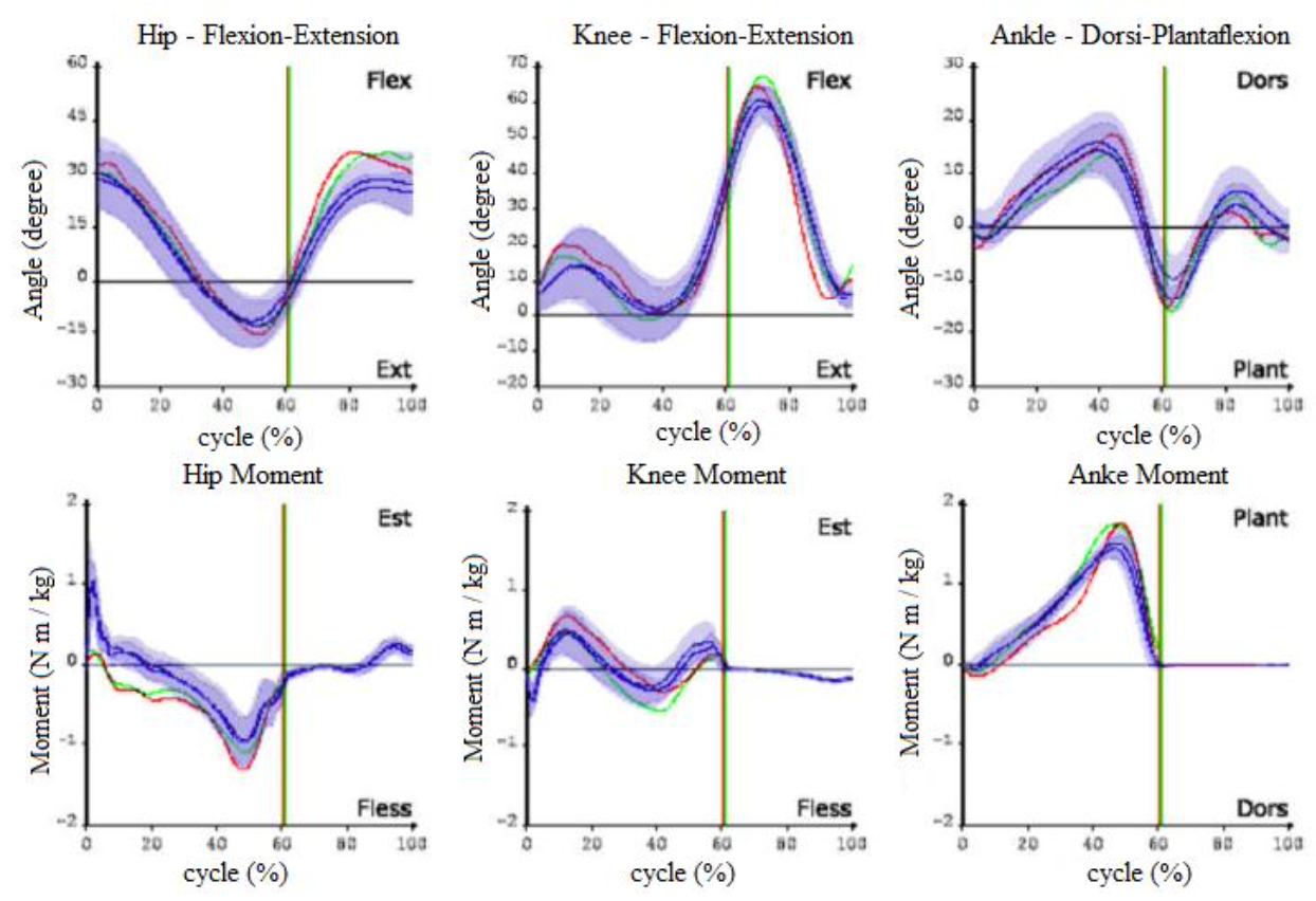
Abstract: In orthopedics, the current internal fixations often use screws or intramedullary rods that obstruct bone material. In this paper, an internal implant was modelled as a hollow cylindrical sector made of a functionally graded material (FGM), which will hold bone in place with less obstruction of bone surface. Functionally graded implant was considered as an inhomogeneous composite structure, with continuously compositional variation from a ceramic at the outer diameter to a metal at the inner diameter. The buckling behavior of the implant was numerically analyzed using a finite element analysis software (ANSYS), and the structural stability of the implant was assessed. The buckling critical loads were calculated for different fixation lengths, cross sectional areas, and different sector angles. These critical loads were then compared with the critical loads of an FGM hollow cylinder with the same cross sectional area. Results showed that the critical load of the hollow cylindrical sector was ∼ 63%, ∼ 70%, and ∼ 73% of the hollow cylinder for different fixation lengths, cross sectional areas, and sector angles, respectively. Further investigations are warranted to study the relation between the composition profile and the implant stability, which can lead to batter internal fixation solutions.
Publication Thumbnail

Taebi, A., Zago, M., Condoluci, C., Galli, M. (2017).
Mechanical work and power analysis in the joints of the lower extremity of adults with Down syndrome during plane walking.
ASME International Mechanical Engineering Conference and Exposition, Tampa, FL.
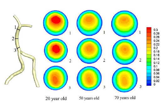
Abstract: Individuals with Down syndrome (DS) use a different motor gait strategy than healthy people. This study aims at analyzing plane walking differences between two groups of normally developed (ND) subjects and subjects with DS in terms of the generated mechanical power and work in the joints of the lower limb. Thirty-nine adults including two groups of 21 subjects with DS (age: 21.6 ± 7 years (mean ± SD)) and 18 ND subjects (age: 25.1 ± 2.4 years) participated in this study. Gait data and ground reaction forces were acquired using a quantitative movement analysis system composed of an optoelectronic motion analyzer (Elite2002, BTS) with eight infrared cameras, and two force platforms mounted in the middle of walkway. Mechanical power and work exchanges were computed during the stance phase by dedicated software, and then compared between the two groups (significance level: p-value = 0.05). Results showed that the mechanical power at the ankle joint was significantly larger in ND subjects compared to subjects with DS (0.084 ± 0.015 vs 0.027 ± 0.010 W/kg). The mechanical work of the ankle joint and the knee joint was significantly lower in ND compared to DS (0.015 ± 0.013 vs 0.028 ± 0.008 kJ/kg.m, and 0.066 ± 0.031 vs 0.109 ± 0.023 kJ/kg.m, respectively). For both groups, the mechanical work done by knee was less than that performed at the ankle and hip level, which might indicate that the knee muscles mainly absorb the energy, rather than generate it. Our results suggest that the subjects with DS walk with a different motor strategy than normal subjects in terms of mechanical power and work in the joints of the lower extremity. Further investigations are warranted to study the relation between these parameters and gait strategy in subjects with DS, which can lead to better rehabilitative strategies.
Publication Thumbnail

Sharifi, A., Salari, A., Taebi, A., Niazmand, H., Niazmand, M.J. (2017).
Flow patterns and wall shear stress distribution in human vertebrobasilar system: A computational study to investigate smoking effects on atherosclerotic stenosis at different ages.
ASME International Mechanical Engineering Conference and Exposition, Tampa, FL.
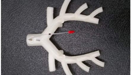
Publication Thumbnail

Azad, M.K., Taebi, A., Mansy, J.H., Mansy, H.A. (2017).
Pressure Loss and Sound Generated in a Miniature Pig Airway Tree Model.
Journal of Applied Biotechnology and Bioengineering, 3(6): 00086.
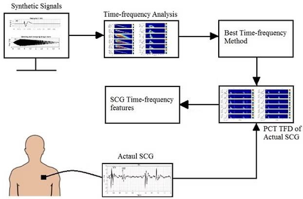
Abstract: Background: Pulmonary auscultation is a common tool for diagnosing various respiratory diseases. Previous studies have documented many details of pulmonary sounds in humans. However, information on sound generation and pressure loss inside animal airways is scarce. Since the morphology of animal airways can be significantly different from human, the characteristics of pulmonary sounds and pressure loss inside animal airways can be different. Objective: The objective of this study is to investigate the sound and static pressure loss measured at the trachea of a miniature pig airway tree model based on the geometric details extracted from physical measurements. Methods: In the current study, static pressure loss and sound generation measured in the trachea was documented at different flow rates of a miniature pig airway tree. Results: Results showed that the static pressure and the amplitude of the recorded sound at the trachea increased as the flow rate increased. The dominant frequency was found to be around 1840-1870Hz for flow rates of 0.2-0.55lit/s. Conclusion: The results suggested that the dominant frequency of the measured sounds remained similar for flow rates from 0.20 to 0.55lit/s. Further investigation is needed to study sound generation under different inlet flow and pulsatile flow conditions.
Publication Thumbnail

Taebi, A., Mansy, H.A. (2017).
Time-frequency Distribution of Seismocardiographic Signals: A Comparative Study.
Bioengineering, 4(2): 32.
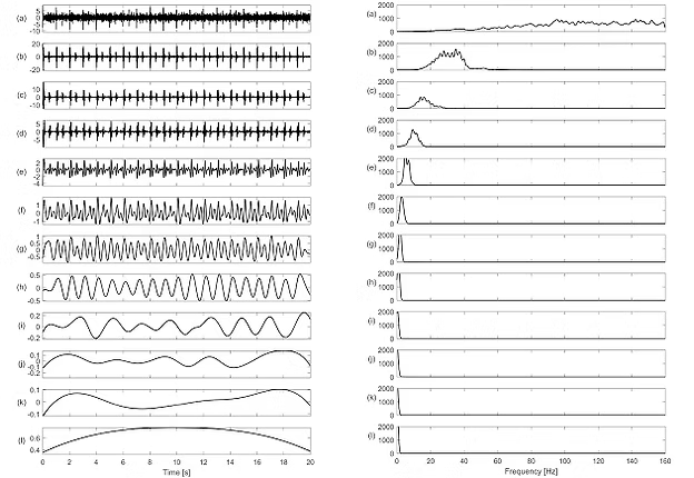
Abstract: Accurate estimation of seismocardiographic (SCG) signal features can help successful signal characterization and classification in health and disease. This may lead to new methods for diagnosing and monitoring heart function. Time-frequency distributions (TFD) were often used to estimate the spectrotemporal signal features. In this study, the performance of different TFDs (e.g., short-time Fourier transform (STFT), polynomial chirplet transform (PCT), and continuous wavelet transform (CWT) with different mother functions) was assessed using simulated signals, and then utilized to analyze actual SCGs. The instantaneous frequency (IF) was determined from TFD and the error in estimating IF was calculated for simulated signals. Results suggested that the lowest IF error depended on the TFD and the test signal. STFT had lower error than CWT methods for most test signals. For a simulated SCG, Morlet CWT more accurately estimated IF than other CWTs, but Morlet did not provide noticeable advantages over STFT or PCT. PCT had the most consistently accurate IF estimations and appeared more suited for estimating IF of actual SCG signals. PCT analysis showed that actual SCGs from eight healthy subjects had multiple spectral peaks at 9.20 ± 0.48, 25.84 ± 0.77, 50.71 ± 1.83 Hz (mean ± SEM). These may prove useful features for SCG characterization and classification.
Publication Thumbnail

Taebi, A., Mansy, H.A. (2017).
Noise Cancellation from Vibrocardiographic Signals Based on the Ensemble Empirical Mode Decomposition.
Journal of Applied Biotechnology and Bioengineering, 2(2): 00024.
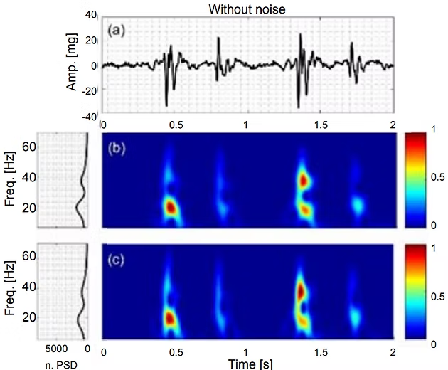
Abstract: Vibrocardiographic (VCG) signals are the cardiac vibration measured at the chest surface. These signals can contain useful information for diagnosing cardiac conditions but are often contaminated by noise. Although band-pass and adaptive filters were used for noise removal from similar signals, the utility of ensemble empirical mode decomposition (EEMD) for filtering VCG was not previously investigated. In this study, an EEMD-based filter was proposed and tested. The filtering scheme first decomposed the VCG waveform into a set of intrinsic mode functions (IMF) then utilized the partial sum of IMFs to remove white noise that was added to simulated VCG signals. To measure the filter effectiveness, the normalized root-mean-square error (NRMSE) between the clean (i.e., before adding noise) and filtered signals was calculated for signal-to-noise ratios ranging from 1 to 20dB. The EEMD-based filter performance was also compared with traditional methods such as Wiener filter. This comparison suggested that EEMD-based filter outperformed the Wiener filter in noise removal from simulated VCG. These results also suggested that EEMD may be utilized for white noise removal from actual VCG signals. Further investigations are warranted to study the relation between IMFs and different types of noise, which can enhance the effectiveness of EEMD-based filters in removing these noise types from actual VCG signals.
Publication Thumbnail

Taebi, A., Mansy, H.A. (2016).
Effect of noise on time-frequency analysis of Vibrocardiographic signals.
Journal of Bioengineering and Biomedical Science, 6(202), 2.
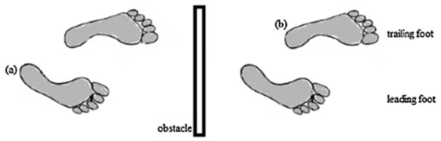
Abstract: Recordings of biological signals such as vibrocardiography often contain contaminating noise. Noise sources may include respiratory, gastrointestinal, and muscles movement, or environmental noise. Depending on individual physiology and sensor location, the vibrocardiographic (VCG) signals may be obscured by these noises in the time-frequency plane, which may interfere with automated characterization of VCG. In this study, polynomial chirplet transform (PCT) and smoothed pseudo Wigner-Ville distribution (SPWVD) were used to estimate the instantaneous frequency (IF) of two simulated VCG signals. One simulated signal contained a time-varying IF while the other had a fixed IF. The error in estimating IF was then calculated for signal-to-noise ratios (SNR) from -10 to 10 dB. Analysis was repeated 100 times at each level of noise using randomized sets of white noise. Error analysis showed that the range of errors in estimating IF was wider when SNR decreased. Results also showed that PCT tended to outperform SPWVD at high SNR. For example, PCT was more accurate at SNR > 3 dB for a simulated VCG signal with constant frequency components, at SNR>-10 dB for a simulated VCG signal with time-varying frequency, and at SNR > 0 for an actual VCG.
Publication Thumbnail

Salami, F., Vimercati, S.L, Rigoldi, C., Taebi, A., Albertini, G., Galli, M. (2014).
Mechanical energy assessment of adults with Down syndrome during walking with obstacle avoidance.
Research in Developmental Disabilities, 35(8), 1856-1862.

Abstract: The aim of this study is analyzing the differences between plane walking and stepping over an obstacle for two groups of healthy people and people with Down syndrome and then, evaluating the movement efficiency between the groups by comprising of their mechanical energy exchanges. 39 adults including two groups of 21 people with Down syndrome (age: 21.6 ± 7 years) and 18 healthy people (age: 25.1 ± 2.4 years) participated in this research. The test has been done in two conditions, first in plane walking and second in walking with an obstacle (10% of the subject's height). The gait data were acquired using quantitative movement analysis, composed of an optoelectronic system (Elite2002, BTS) with eight infrared cameras. Mechanical energy exchanges are computed by dedicated software and finally the data including spatiotemporal parameters, mechanical energy parameters and energy recovery of gait cycle are analyzed by statistical software to find significant differences. Regards to spatiotemporal parameters velocity and step length are lower in people with Down syndrome. Mechanical energy parameters particularly energy recovery does not change from healthy people to people with Down syndrome. However, there are some differences in inter-group through plane walking to obstacle avoidance and it means people with Down syndrome probably use their residual abilities in the most efficient way to achieve the main goal of an efficient energy recovery.

Publication Statistics