

Read Dr. Cassimeris' recently published research findings
Equine Laminitis: Cellular and Molecular Mechanisms Underlying Disease
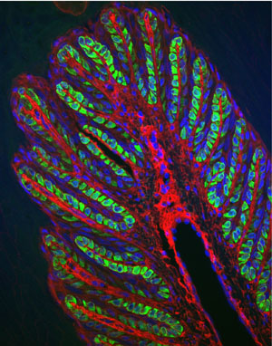
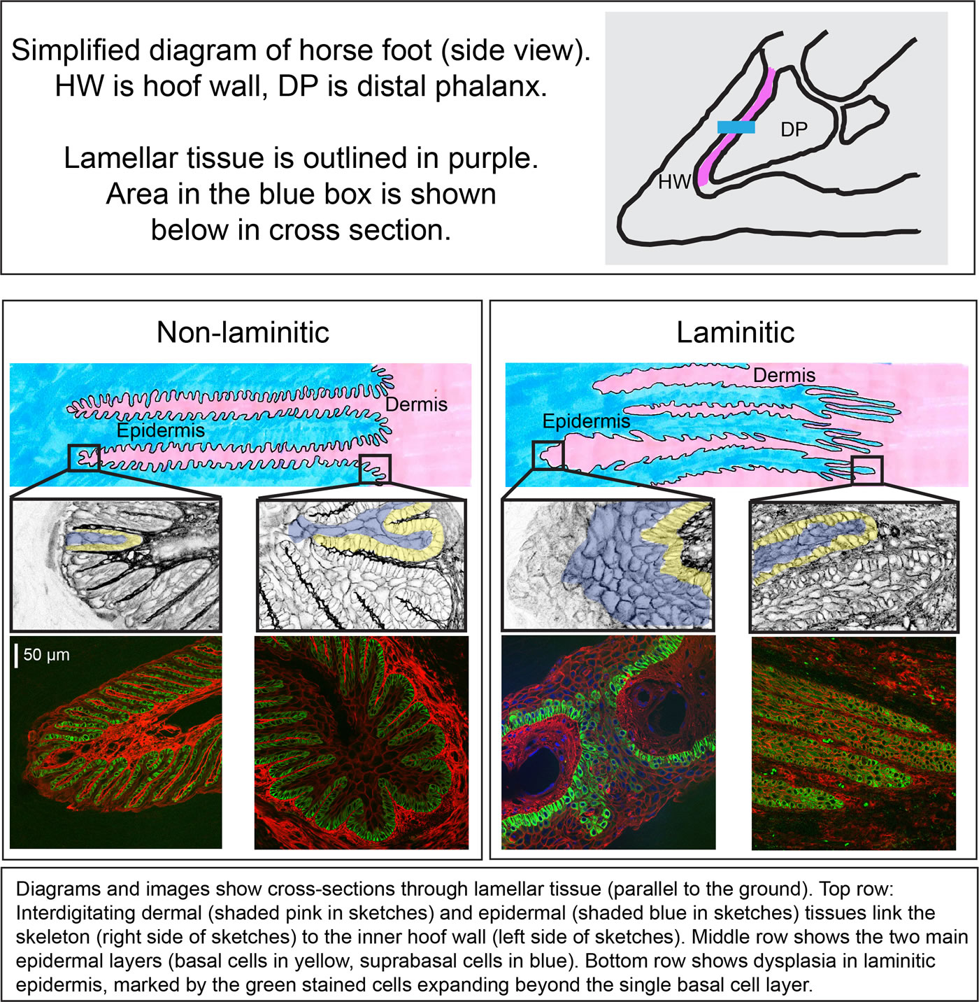
Laminitis is a painful disease in horses and the 2nd leading cause of euthanasia. Laminitis results when the lamellae, which normally connect the hoof wall and the skeleton, are compromised and lose function. The lamellae are derived from skin and are folded to increase the surface area attaching epidermal tissues on the hoof side and dermal tissues on the skeletal side. These tissues are diagramed in the figure.
What do we want to investigate over the next year or two?
Our broad goals are: (1) to uncover the cellular and molecular changes responsible for disease, and (2) to link mechanisms underlying laminitis with those causing several human diseases, which will provide insight into disease progression and suggest therapeutic options.
Earlier studies lead to the development of two main research goals. We expect that each project will reveal steps in the disease process, and that together the two projects will provide a better understanding of why the lamellar tissue is compromised in disease and how best to interrupt or prevent these changes.
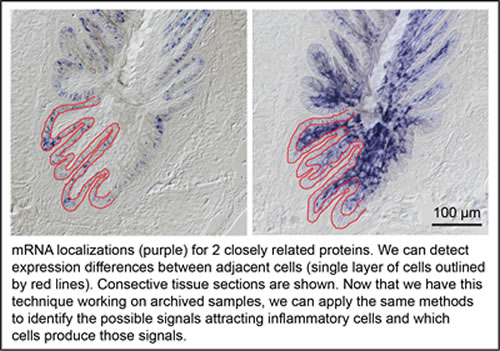
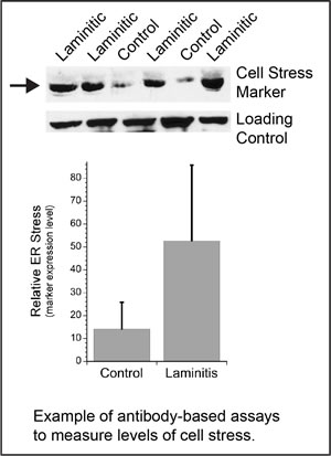 Project 1 continues research to identify cellular stress pathways active in endocrinopathy-triggered laminitis (including equine metabolic syndrome and PPID/Cushings disease) and supporting limb laminitis. Initial data highlight two stress pathways that are also active in many human diseases including type 2 diabetes and neurodegenerative diseases such as Alzheimer's and Parkinson's diseases. Over the last 2 years, we discovered tools suitable for the investigation of two stress pathways in equine tissues and found activation of these two stress pathways in chronic laminitis (some of our data is shown in the figures, a manuscript describing our results is currently in peer review for publication). Next, we need to look at earlier, acute states. We need to know whether cellular stress is causing an active cell death process (apoptosis), and/or whether these stress pathways are causing abnormal characteristics in epidermal cells and loss of their normal functions to maintain the structure of the lamellae. Either outcome can damage lamellar epidermal tissue and lead to inflammation (Project 2). Project 1 dovetails with on-going research in Dr. Galantino-Homer's lab.
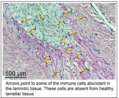
Project 2 will uncover the inflammatory pathways active in endocrinopathic and supporting limb laminitis, similar to the "sterile" inflammation observed in many human diseases. These inflammatory pathways are activated in response to tissue damage caused by cell stress (examined in Project 1). A large number of immune cells (leukocytes and lymphocytes) are present in laminitic tissue, indicating that signals are calling these cells into the lamellae. We want to know what these signal(s) are and which lamellar cells produce them. We expect that stressed epidermal cells raise the alarm and call in cells of the immune system; the resulting inflammation and tissue damage then begins a domino effect, causing more cell loss, and disease. We have candidate molecules for the signal(s) and tools to identify them. If pro-inflammatory mediators, the "come hither" signals, are similar to those in human disease, therapeutic options would be available. The latter is a long-term goal that will not be addressed in the proposed study.
 |
 |
Where will we get samples for study? Samples are available from archived tissues provided by Dr. Galantino-Homer and the Laminitis Discovery Database, New Bolton Center, Univ. Penn. School of Veterinary Medicine. Tissues were collected from horses undergoing euthanasia and donated for research.
All research is conducted in accordance with local, state and federal guidelines.

Lynne Cassimeris, Ph.D.
Professor
Department of Biological Sciences
Lehigh University
Hannah Galantino-Homer, VMD, Ph.D.
Senior Research
Investigator
New Bolton Center
University of Pennsylvania

Julie Engiles, VMD
Associate Professor of Pathology
Department of Pathobiology-New Bolton Center
University of Pennsylvania
For the past 3 years, we have collaborated with Dr. Galantino-Homer and colleagues at the University of Pennsylvania School of Veterinary Medicine and applied our nearly 35 years of research experience in cell and molecular biology to understand the cellular processes underlying tissue failure in laminitis. In addition to Dr. Galantino-Homer, we collaborate closely with Dr. Engiles, a board-certified veterinary pathologist. More recently, Dr. van Eps, who obtained his PhD in equine laminitis under Dr. Pollitt of the University of Queensland, Queensland, Australia and who is a board-certified specialist in equine internal medicine, has joined the faculty at New Bolton Center and is beginning to collaborate with us on these projects. Our initial results drive the next research goals, described in "Research Projects."
Who will do the actual work and why do we need money?
Lynne Cassimeris, Ph.D. will lead a research group at Lehigh University and collaborate with Dr. Galantino-Homer, DMD, Ph.D., a laminitis expert, and Julie Engiles, VMD, a veterinary pathologist.
Dr. Cassimeris donates her time and does not receive any salary compensation. Drs. Galantino-Homer and Engiles are supported by the University of Pennsylvania and also will not require salary support for this work.
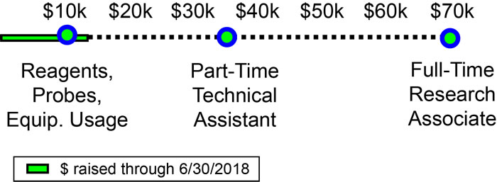
Funds are needed to design, develop and validate biological reagents for the research projects. We have available shared infrastructure and equipment needed for the proposed work and will not need to purchase any equipment, but we need funds to cover user fees associated with maintenance of high end equipment. Ideally, to speed research progress, financial support will allow us to hire a post-doctoral fellow or lab technician to perform the day-to-day experiments. A small Faculty Incentive Grant from Lehigh University allowed Dr. Cassimeris to hire an outstanding post-doc; even at part time effort, Dr. Da Silva Santos acquired data contributing to 3 manuscripts that are currently in preparation. Undergraduates will also participate in the research projects. Caitlyn Wilson (now in her first year at UC Davis School of Veterinary Medicine) is a co-author on a manuscript in preparation and has already co-authored two abstracts, one presented at a national conference and another at a local diabetes meeting.
Long term, we hope to be competitive for external support either from foundations or through federal grants from the National Institutes of Health (NIH) or the National Science Foundation (NSF). Dr. Cassimeris has been funded by NIH almost continuously during her 26 years directing an independent lab group at Lehigh. She has also received funding from NSF, as well as several other federal agencies.
Donate by check (payable to Lehigh University) and mailed to:
Laminitis Project
Lehigh University - Dept. of Biological Sciences
111 Research Dr.
Bethlehem, PA 18015
- or -
Click here to donate via Lehigh's Development Office.
Then enter information as outlined below.
Please specify that donation is for the LAMINITIS PROJECT.

Click here for a PDF of these instructions.
Please be sure to specify under “other” that you are donating to the Laminitis Project.