About JFilament
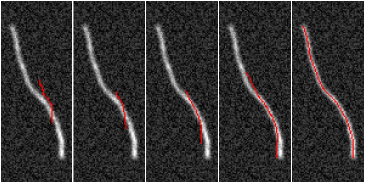
JFilament is an ImageJ/Fiji plugin for segmentation and tracking of 2D and 3D filaments in fluorescenece microscopy images [1]. It allows users to segment linear elements and track them in time.
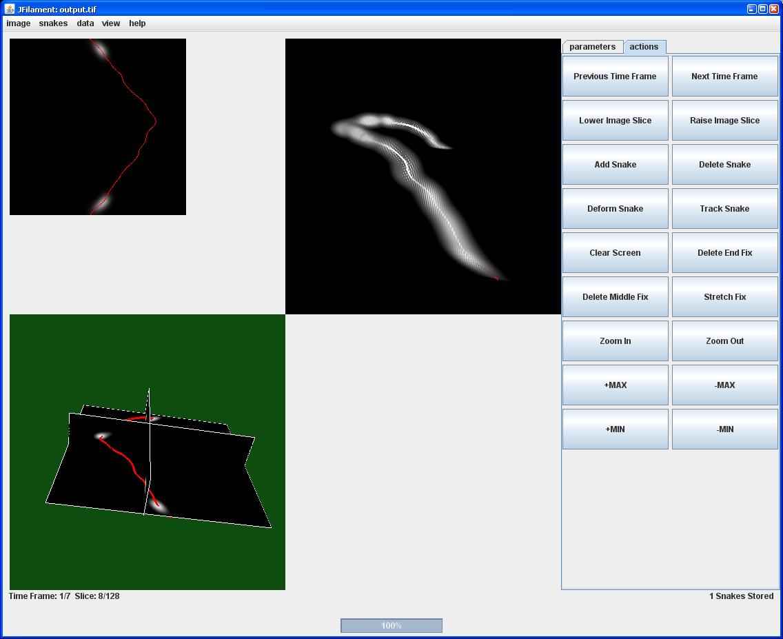
The method is based on stretching open active contours or "snakes" [2] that deform to bright linear elements in an image. Users can deform, add, delete, save and load filament curves. Images can be visualized together with graphical curves representing segmented filaments.
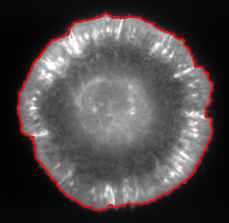
JFilament also includes 2D "closed" active contours which can be used for tasks such as segmentation and tracking of cell boundaries.
The software is free to use and modify by referencing [1]. The current release is maintained by Matthew Smith.
JFilament is optimized for semi-automated tracking of multiple filaments with ability of manual edit. For networks and more automated methods, see SOAX (for static 2D/3D images) and TSOAX (includes 2D/3D tracking) software.
Platform
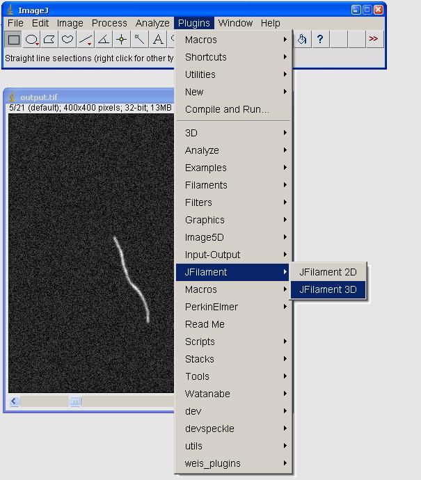
JFilament is an ImageJ / Fiji plugin consisting of two components: JFilament3D and JFilament2D, optimized for 3D and 2D images, respectively. JFilament3D has the prerequisite of Java3D for 3D visualization.
JFilament can be easily installed in Fiji from update site http://sites.imagej.net/Odinsbane/ This can be added in Help, Update Fiji.
It can also be downloaded and installed in the ImageJ plugins directory, as described in the downloads page.
References
This work was supported by NIH, grant R21GM083928 and by the Biosystems Dynamics Summer Institute at Lehigh University, funded by the Howard Hughes Medical Institute.
[1] M. B. Smith, H. Li, T. Shen, X. Huang, E. Yusuf, and D. Vavylonis, "Segmentation and Tracking of Cytoskeletal Filaments using Open Active Contours," Cytoskeleton (2010) DOI: 10.1002/cm.20481.
[2] H. Li, T. Shen, M. B. Smith, I. Fujiwara, D. Vavylonis, and X. Huang, "Automated Actin Filament Segmentation, Tracking, and Tip Elongation Measurements based on Open Active Contour Models," In Proc. of the IEEE International Symposium on Biomedical Imaging: From Nano to Macro, ISBI-09, Boston, 2009

Department of Physics, Lehigh University
Department of Computer Science and Engineering, Lehigh University