| Physics Department | Center For Optical Technologies | Lehigh University |

|
Triplet exciton diffusion in rubrene![imaging triplet diffusion [Image: triplet diffusion]](stor/imagingtripletdiffusion.jpg)
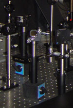
Setup for triplet exciton microscopy. Blue light is focused on the surface of the crystal, where photoexcited singlet states undergo fission to create a population of long lived triplet excitons that visibly diffuse. ![imaging triplet diffusion [Image: triplet diffusion]](stor/excitondiffusion.jpg)
Excitation and fluorescence intensity patterns when illuminating a natural facet of a rubrene crystal that is perpendicular to the c-axis of the crystal. The high efficiency of singlet exciton fission, the absence of significant triplet exciton annihilation that doesn't occur via fusion and photon emission, and the large triplet exciton lifetime of about 100 microseconds combine to allow for triplet exciton imaging. Yes, triplet excitons are dark states, but in rubrene they can collide with each other, and such a collision event can lead to fusion, the formation of a singlet state, and then photon emission. This enables experiments like that shown in the figure to the right. A blue photoexcitation laser can be tightly focused on a rubrene surface while the surface is imaged via confocal imaging. By then changing transmission filters in the path that leads from sample to detection camera, it is possible to either image the surface under illumination from the excitation light, or else one can image only the red fluorescence. Under high magnification (use a 100x objective) on the large facet (normally the facet perpendicular to the c-axis of rubrene) of a crystal one can then see that that the image obtained using the excitation light is what one would expect, the image of a focused beam. But the image obtained using the fluorescence light has a different shape. The luminescence extends much further than the excitation light, in particular along the b-axis of the crystal, which corresponds to the close stacking direction of the rubrene molecules. ![imaging triplet diffusion [Image: triplet diffusion]](stor/rubrenetripletdiffusion.jpg)
Determination of the triplet exciton diffusion length in rubrene. While straight imaging on a linear scale, even when plotted as a fake color plot, essentially just shows that the triplet exciton population has expanded along a specific preferred direction, plotting the fluorescence intensity as a function of position shows that the fluorescence decreases exponentially as one moves further away from the excitation spot. This is exactly what is predicted by the diffusion equation for particles with a finite lifetime, which predicts an exponential decay in the density of the particle with an exponential constant that can be assigned to the diffusion length. When imaging fluorescence induced by triplet fusion, one must then take into account that the fluorescence is proportional to the square of the triplet exciton density, which then means that the actual average diffusion length of a triplet exciton is twice the exponential decay time of the fluorescence intensity. To demonstrate that what is observed is really the diffusion of triplet excitons it is possible to study the variability of the fluorescence intensity pattern as one illuminates different facets of a rubrene crystal, something that becomes possible if one decides to harvest the small sub-mm chubby crystals that are not good for doing experiments that requires transmitting light through them, but are very good when it comes to finding a facet that is not the usual large facet of as grown crystal platelets. ![singlets and triplets [Image: excitons]](stor/diffusionpatterns.jpg)
Different fluorescence patterns obtained under different conditions as the triplet excitons diffuse in two crystals at the same time (A), in a single crystal with the high mobility axis horizontal (B) or on a crystal facet for which the high mobility direction is not parallel to the surface, but instead is pointing downwards into the crystal. More information:
See also:
|
| Contact | Goto Top of Page |